 Movie
Movie Controller

[English] 日本語
 Yorodumi
- PDB-3j42: Obstruction of Dengue Virus Maturation by Fab Fragments of the 2H... -
+ Open data

- Basic information
| Entry | Database: PDB / ID: 3j42 | ||||||
|---|---|---|---|---|---|---|---|
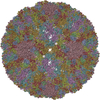
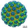
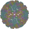
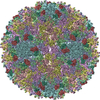
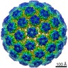
| Title | Obstruction of Dengue Virus Maturation by Fab Fragments of the 2H2 Antibody | ||||||
 Components Components |
| ||||||
 Keywords Keywords | VIRUS/IMMUNE SYSTEM / Dengue / maturation / immature / antibody / VIRUS-IMMUNE SYSTEM complex | ||||||
| Function / homology |  Function and homology information Function and homology informationsymbiont-mediated suppression of host JAK-STAT cascade via inhibition of host TYK2 activity / host cell mitochondrion / immunoglobulin complex / symbiont-mediated suppression of host JAK-STAT cascade via inhibition of STAT2 activity / symbiont-mediated suppression of host cytoplasmic pattern recognition receptor signaling pathway via inhibition of MAVS activity / viral capsid / ribonucleoside triphosphate phosphatase activity / double-stranded RNA binding / channel activity / monoatomic ion transmembrane transport ...symbiont-mediated suppression of host JAK-STAT cascade via inhibition of host TYK2 activity / host cell mitochondrion / immunoglobulin complex / symbiont-mediated suppression of host JAK-STAT cascade via inhibition of STAT2 activity / symbiont-mediated suppression of host cytoplasmic pattern recognition receptor signaling pathway via inhibition of MAVS activity / viral capsid / ribonucleoside triphosphate phosphatase activity / double-stranded RNA binding / channel activity / monoatomic ion transmembrane transport / clathrin-dependent endocytosis of virus by host cell / molecular adaptor activity / adaptive immune response / methyltransferase cap1 activity / mRNA 5'-cap (guanine-N7-)-methyltransferase activity / RNA helicase activity / protein dimerization activity / symbiont-mediated suppression of host innate immune response / immune response / host cell perinuclear region of cytoplasm / host cell endoplasmic reticulum membrane / symbiont-mediated suppression of host type I interferon-mediated signaling pathway / serine-type endopeptidase activity / symbiont-mediated activation of host autophagy / viral RNA genome replication / RNA-directed RNA polymerase activity / fusion of virus membrane with host endosome membrane / viral envelope / lipid binding / virion attachment to host cell / host cell nucleus / virion membrane / structural molecule activity / proteolysis / extracellular region / ATP binding / metal ion binding / plasma membrane Similarity search - Function | ||||||
| Biological species |   Dengue virus 2 Dengue virus 2 | ||||||
| Method | ELECTRON MICROSCOPY / single particle reconstruction / cryo EM / Resolution: 21 Å | ||||||
 Authors Authors | Wang, Z. / Pennington, J.G. / Jiang, W. / Rossmann, M.G. | ||||||
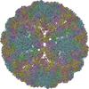
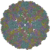
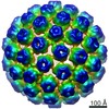
 Citation Citation | Journal: J Virol / Year: 2013 Title: Obstruction of dengue virus maturation by Fab fragments of the 2H2 antibody. Authors: Zhiqing Wang / Long Li / Janice G Pennington / Ju Sheng / Moh Lan Yap / Pavel Plevka / Geng Meng / Lei Sun / Wen Jiang / Michael G Rossmann /  Abstract: The 2H2 monoclonal antibody recognizes the precursor peptide on immature dengue virus and might therefore be a useful tool for investigating the conformational change that occurs when the immature ...The 2H2 monoclonal antibody recognizes the precursor peptide on immature dengue virus and might therefore be a useful tool for investigating the conformational change that occurs when the immature virus enters an acidic environment. During dengue virus maturation, spiky, immature, noninfectious virions change their structure to form smooth-surfaced particles in the slightly acidic environment of the trans-Golgi network, thereby allowing cellular furin to cleave the precursor-membrane proteins. The dengue virions become fully infectious when they release the cleaved precursor peptide upon reaching the neutral-pH environment of the extracellular space. Here we report on the cryo-electron microscopy structures of the immature virus complexed with the 2H2 antigen binding fragments (Fab) at different concentrations and under various pH conditions. At neutral pH and a high concentration of Fab molecules, three Fab molecules bind to three precursor-membrane proteins on each spike of the immature virus. However, at a low concentration of Fab molecules and pH 7.0, only two Fab molecules bind to each spike. Changing to a slightly acidic pH caused no detectable change of structure for the sample with a high Fab concentration but caused severe structural damage to the low-concentration sample. Therefore, the 2H2 Fab inhibits the maturation process of immature dengue virus when Fab molecules are present at a high concentration, because the three Fab molecules on each spike hold the precursor-membrane molecules together, thereby inhibiting the normal conformational change that occurs during maturation. | ||||||
| History |
|
- Structure visualization
| Movie |
Movie viewer |
|---|---|
| Structure viewer | Molecule: MolmilJmol/JSmol |
- Downloads & links
-Download
| PDBx/mmCIF format | 3j42.cif.gz | 291.2 KB | Display | PDBx/mmCIF format |
|---|---|---|---|---|
| PDB format | pdb3j42.ent.gz | 229.5 KB | Display | PDB format |
| PDBx/mmJSON format | 3j42.json.gz | Tree view | PDBx/mmJSON format | |
| Others |  Other downloads Other downloads |
-Validation report
| Arichive directory | https://data.pdbj.org/pub/pdb/validation_reports/j4/3j42ftp://data.pdbj.org/pub/pdb/validation_reports/j4/3j42 | HTTPS FTP |
|---|
-Related structure data
| Related structure data | 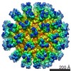 5674MC 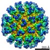 5675C 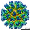 5676C 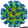 5677C 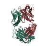 4kvcC M: map data used to model this data C: citing same article ( |
|---|---|
| Similar structure data |

-Links
 PDBj
PDBj


- Assembly
| Deposited unit | 
|
|---|---|
| 1 | x 60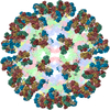
|
| 2 |
|
| 3 | x 5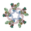
|
| 4 | x 6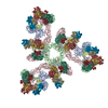
|
| 5 | 
|
| Symmetry | Point symmetry: (Schoenflies symbol: I (icosahedral)) |
-Components
| #1: Protein | Mass: 43400.898 Da / Num. of mol.: 3 / Fragment: UNP residues 281-675 / Source method: isolated from a natural source / Source: (natural) Dengue virus 2 / References: UniProt: O11875#2: Protein | Mass: 9261.531 Da / Num. of mol.: 3 / Fragment: UNP residues 1-81 / Source method: isolated from a natural source / Source: (natural) Dengue virus 2 / References: UniProt: Q3BCY5#3: Antibody | Mass: 23696.553 Da / Num. of mol.: 3 / Fragment: SEE REMARK 999 Source method: isolated from a genetically manipulated source Source: (gene. exp.) #4: Antibody | Mass: 23355.746 Da / Num. of mol.: 3 / Fragment: SEE REMARK 999 Source method: isolated from a genetically manipulated source Source: (gene. exp.) Has protein modification | Y | Sequence details | 2H2 HEAVY CHAIN FAB IS A CHIMERA COMPRISING RESIDUES 17-119 OF UNP P01783 AND RESIDUES 120-230 OF ...2H2 HEAVY CHAIN FAB IS A CHIMERA COMPRISING | |
|---|
-Experimental details
-Experiment
| Experiment | Method: ELECTRON MICROSCOPY |
|---|---|
| EM experiment | Aggregation state: PARTICLE / 3D reconstruction method: single particle reconstruction |
- Sample preparation
| Component | Name: High-concentration Fab Fragment of 2H2 antibody binding to immature Dengue virus at pH 7 Type: VIRUS Details: Three Fab fragments bind to three pr on each trimeric spike |
|---|---|
| Details of virus | Empty: NO / Enveloped: YES / Host category: INVERTEBRATE / Isolate: STRAIN / Type: VIRION |
| Natural host | Organism: Aedes albopictus / Strain: C6-36 |
| Buffer solution | Name: 100 mM phosphate buffer / pH: 7 / Details: 100 mM phosphate buffer |
| Specimen | Embedding applied: NO / Shadowing applied: NO / Staining applied: NO / Vitrification applied: YES |
| Specimen support | Details: Quantifoil 200 mesh |
| Vitrification | Instrument: HOMEMADE PLUNGER / Cryogen name: ETHANE / Temp: 100 K / Details: Plunged into liquid ethane (homemade plunger) |
- Electron microscopy imaging
| Microscopy | Model: FEI/PHILIPS CM200FEG / Date: Apr 7, 2011 |
|---|---|
| Electron gun | Electron source:  FIELD EMISSION GUN / Accelerating voltage: 200 kV / Illumination mode: FLOOD BEAM FIELD EMISSION GUN / Accelerating voltage: 200 kV / Illumination mode: FLOOD BEAM |
| Electron lens | Mode: BRIGHT FIELD / Nominal magnification: 50000 X / Calibrated magnification: 51040 X / Nominal defocus max: 5000 nm / Nominal defocus min: 2000 nm / Cs: 2 mm Astigmatism: Objective lens astigmatism was corrected at 100,000 times magnification Camera length: 0 mm |
| Specimen holder | Specimen holder model: GATAN LIQUID NITROGEN / Temperature: 100 K / Temperature (max): 105 K / Temperature (min): 80 K / Tilt angle max: 0 ° / Tilt angle min: 0 ° |
| Image recording | Electron dose: 25 e/Å2 / Film or detector model: KODAK SO-163 FILM |
| Image scans | Num. digital images: 49 |
| Radiation | Protocol: SINGLE WAVELENGTH / Monochromatic (M) / Laue (L): M / Scattering type: x-ray |
| Radiation wavelength | Relative weight: 1 |
- Processing
| EM software |
| |||||||||||||||||||||
|---|---|---|---|---|---|---|---|---|---|---|---|---|---|---|---|---|---|---|---|---|---|---|
| CTF correction | Details: Film | |||||||||||||||||||||
| Symmetry | Point symmetry: I (icosahedral) | |||||||||||||||||||||
| 3D reconstruction | Method: projection matching / Resolution: 21 Å / Resolution method: FSC 0.5 CUT-OFF / Num. of particles: 378 / Nominal pixel size: 3.72 Å / Actual pixel size: 3.72 Å / Details: (Single particle--Applied symmetry: I) / Symmetry type: POINT | |||||||||||||||||||||
| Atomic model building | Protocol: RIGID BODY FIT / Space: REAL / Details: REFINEMENT PROTOCOL--rigid body | |||||||||||||||||||||
| Atomic model building |
| |||||||||||||||||||||
| Refinement step | Cycle: LAST
|
