 Movie
Movie Controller

[English] 日本語
 Yorodumi
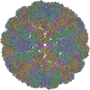
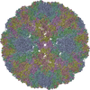
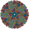
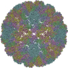
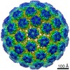
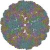
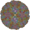
- PDB-3jba: The U4 antibody epitope on human papillomavirus 16 identified by ... -
+ Open data

- Basic information
| Entry | Database: PDB / ID: 3jba | ||||||
|---|---|---|---|---|---|---|---|
| Title | The U4 antibody epitope on human papillomavirus 16 identified by cryo-EM | ||||||
 Components Components |
| ||||||
 Keywords Keywords | VIRUS/IMMUNE SYSTEM / HPV16 / antibody / U4 / neutralization / Fab / VIRUS-IMMUNE SYSTEM complex | ||||||
| Function / homology |  Function and homology information Function and homology informationT=7 icosahedral viral capsid / endocytosis involved in viral entry into host cell / virion attachment to host cell / host cell nucleus / structural molecule activity Similarity search - Function | ||||||
| Biological species |  Human papillomavirus type 16 Human papillomavirus type 16 | ||||||
| Method | ELECTRON MICROSCOPY / single particle reconstruction / cryo EM / Resolution: 12 Å | ||||||
 Authors Authors | Guan, J. / Bywaters, S.M. / Brendle, S.A. / Lee, H. / Ashley, R.E. / Christensen, N.D. / Hafenstein, S. | ||||||
 Citation Citation | Journal: J Virol / Year: 2015 Title: The U4 Antibody Epitope on Human Papillomavirus 16 Identified by Cryo-electron Microscopy. Authors: Jian Guan / Stephanie M Bywaters / Sarah A Brendle / Hyunwook Lee / Robert E Ashley / Neil D Christensen / Susan Hafenstein /  Abstract: The human papillomavirus (HPV) major structural protein L1 composes capsomers that are linked together through interactions mediated by the L1 C terminus to constitute a T=7 icosahedral capsid. H16. ...The human papillomavirus (HPV) major structural protein L1 composes capsomers that are linked together through interactions mediated by the L1 C terminus to constitute a T=7 icosahedral capsid. H16.U4 is a type-specific monoclonal antibody recognizing a conformation-dependent neutralizing epitope of HPV thought to include the L1 protein C terminus. The structure of human papillomavirus 16 (HPV16) complexed with H16.U4 fragments of antibody (Fab) was solved by cryo-electron microscopy (cryo-EM) image reconstruction. Atomic structures of virus and Fab were fitted into the corresponding cryo-EM densities to identify the antigenic epitope. The antibody footprint mapped predominately to the L1 C-terminal arm with an additional contact point on the side of the capsomer. This footprint describes an epitope that is presented capsid-wide. However, although the H16.U4 epitope suggests the presence of 360 potential binding sites exposed in the capsid valley between each capsomer, H16.U4 Fab bound only to epitopes located around the icosahedral five-fold vertex of the capsid. Thus, the binding characteristics of H16.U4 defined in this study showed a distinctive selectivity for local conformation-dependent interactions with specific L1 invading arms between five-fold related capsomers. IMPORTANCE: Human papillomavirus 16 (HPV16) is the most prevalent oncogenic genotype in HPV-associated anogenital and oral cancers. Here we use cryo-EM reconstruction techniques to solve the ...IMPORTANCE: Human papillomavirus 16 (HPV16) is the most prevalent oncogenic genotype in HPV-associated anogenital and oral cancers. Here we use cryo-EM reconstruction techniques to solve the structures of the HPV16 capsid complexes using H16.U4 fragment of antibody (Fab). Different from most other antibodies directed against surface loops, H16.U4 monoclonal antibody is unique in targeting the C-terminal arm of the L1 protein. This monoclonal antibody (MAb) is used throughout the HPV research community in HPV serological and vaccine development and to define mechanisms of HPV uptake. The unique binding mode of H16.U4 defined here shows important conformation-dependent interactions within the HPV16 capsid. By targeting an important structural and conformational epitope, H16.U4 may identify subtle conformational changes in different maturation stages of the HPV capsid and provide a key probe to analyze the mechanisms of HPV uptake during the early stages of virus infection. Our analyses precisely define important conformational epitopes on HPV16 capsids that are key targets for successful HPV prophylactic vaccines. | ||||||
| History |
|
- Structure visualization
| Movie |
Movie viewer |
|---|---|
| Structure viewer | Molecule: MolmilJmol/JSmol |
 UCSF Chimera
- Downloads & links
-Download
| PDBx/mmCIF format | 3jba.cif.gz | 541.7 KB | Display | PDBx/mmCIF format |
|---|---|---|---|---|
| PDB format | pdb3jba.ent.gz | 359.9 KB | Display | PDB format |
| PDBx/mmJSON format | 3jba.json.gz | Tree view | PDBx/mmJSON format | |
| Others |  Other downloads Other downloads |
-Validation report
| Arichive directory | https://data.pdbj.org/pub/pdb/validation_reports/jb/3jbaftp://data.pdbj.org/pub/pdb/validation_reports/jb/3jba | HTTPS FTP |
|---|
-Related structure data
| Related structure data |  6424MC 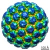 6423C M: map data used to model this data C: citing same article ( |
|---|---|
| Similar structure data |
-Links
 PDBj
PDBj


- Assembly
| Deposited unit | 
|
|---|---|
| 1 | x 60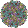
|
| 2 |
|
| 3 | x 5
|
| 4 | x 6
|
| 5 | 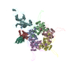
|
| Symmetry | Point symmetry: (Schoenflies symbol: I (icosahedral)) |
-Components
| #1: Antibody | Mass: 12699.254 Da / Num. of mol.: 1 / Fragment: Fab variable domain / Source method: isolated from a natural source / Source: (natural) | ||
|---|---|---|---|
| #2: Antibody | Mass: 12711.025 Da / Num. of mol.: 1 / Fragment: Fab variable domain / Source method: isolated from a natural source / Source: (natural) | ||
| #3: Protein | Mass: 53445.617 Da / Num. of mol.: 6 Source method: isolated from a genetically manipulated source Source: (gene. exp.) Human papillomavirus type 16 / Gene: L1 / Plasmid: SV40 / Cell line (production host): HEK293TT / Production host:  Homo sapiens (human) / References: UniProt: C9E771, UniProt: P03101*PLUS Homo sapiens (human) / References: UniProt: C9E771, UniProt: P03101*PLUSHas protein modification | Y | |
-Experimental details
-Experiment
| Experiment | Method: ELECTRON MICROSCOPY |
|---|---|
| EM experiment | Aggregation state: PARTICLE / 3D reconstruction method: single particle reconstruction |
- Sample preparation
| Component |
| ||||||||||||||||
|---|---|---|---|---|---|---|---|---|---|---|---|---|---|---|---|---|---|
| Molecular weight | Value: 32.8 MDa / Experimental value: NO | ||||||||||||||||
| Details of virus | Empty: NO / Enveloped: NO / Host category: VERTEBRATES / Isolate: SEROTYPE / Type: VIRUS-LIKE PARTICLE | ||||||||||||||||
| Natural host | Organism: Homo sapiens | ||||||||||||||||
| Buffer solution | Name: 1 M NaCl, 200 mM Tris / pH: 7.4 / Details: 1 M NaCl, 200 mM Tris | ||||||||||||||||
| Specimen | Conc.: 1.2 mg/ml / Embedding applied: NO / Shadowing applied: NO / Staining applied: NO / Vitrification applied: YES | ||||||||||||||||
| Specimen support | Details: glow-discharged holey carbon support grid | ||||||||||||||||
| Vitrification | Instrument: GATAN CRYOPLUNGE 3 / Cryogen name: ETHANE / Temp: 102 K / Humidity: 90 % / Details: Plunged into liquid ethane (GATAN CRYOPLUNGE 3). |
- Electron microscopy imaging
| Microscopy | Model: JEOL 2100 / Date: Jul 22, 2014 |
|---|---|
| Electron gun | Electron source: LAB6 / Accelerating voltage: 200 kV / Illumination mode: SPOT SCAN |
| Electron lens | Mode: BRIGHT FIELD / Nominal magnification: 50000 X / Nominal defocus max: 5430 nm / Nominal defocus min: 1600 nm / Cs: 2 mm |
| Specimen holder | Specimen holder model: GATAN LIQUID NITROGEN / Temperature: 95 K |
| Image recording | Electron dose: 15 e/Å2 / Film or detector model: GATAN ULTRASCAN 4000 (4k x 4k) |
| Image scans | Num. digital images: 151 |
| Radiation | Protocol: SINGLE WAVELENGTH / Monochromatic (M) / Laue (L): M / Scattering type: x-ray |
| Radiation wavelength | Relative weight: 1 |
- Processing
| EM software |
| ||||||||||||||||||||||||
|---|---|---|---|---|---|---|---|---|---|---|---|---|---|---|---|---|---|---|---|---|---|---|---|---|---|
| CTF correction | Details: each particle | ||||||||||||||||||||||||
| Symmetry | Point symmetry: I (icosahedral) | ||||||||||||||||||||||||
| 3D reconstruction | Method: Cross-common lines / Resolution: 12 Å / Resolution method: FSC 0.5 CUT-OFF / Num. of particles: 5806 / Nominal pixel size: 2.33 Å / Actual pixel size: 2.33 Å Details: (Single particle details: The particles were selected using semi-automatic program e2boxer.py.) (Single particle--Applied symmetry: I) Symmetry type: POINT | ||||||||||||||||||||||||
| Atomic model building | Protocol: RIGID BODY FIT / Space: REAL / Details: REFINEMENT PROTOCOL--rigid body | ||||||||||||||||||||||||
| Atomic model building | PDB-ID: 3J6R Pdb chain-ID: F / Accession code: 3J6R / Source name: PDB / Type: experimental model | ||||||||||||||||||||||||
| Refinement step | Cycle: LAST
|

