 Movie
Movie Controller

[English] 日本語
 Yorodumi
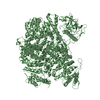
- PDB-3j0a: Homology model of human Toll-like receptor 5 fitted into an elect... -
+ Open data

- Basic information
| Entry | Database: PDB / ID: 3j0a | |||||||||
|---|---|---|---|---|---|---|---|---|---|---|
| Title | Homology model of human Toll-like receptor 5 fitted into an electron microscopy single particle reconstruction | |||||||||
 Components Components | Toll-like receptor 5 | |||||||||
 Keywords Keywords | IMMUNE SYSTEM / Toll-like receptor 5 / membrane protein / leucine-rich repeat / asymmetric homodimer / glycoprotein | |||||||||
| Function / homology |  Function and homology information Function and homology informationToll Like Receptor 5 (TLR5) Cascade / MyD88 deficiency (TLR5) / toll-like receptor 5 signaling pathway / IRAK4 deficiency (TLR5) / MyD88 cascade initiated on plasma membrane / interleukin-1 receptor binding / toll-like receptor signaling pathway / pattern recognition receptor activity / positive regulation of interleukin-8 production / cellular response to mechanical stimulus ...Toll Like Receptor 5 (TLR5) Cascade / MyD88 deficiency (TLR5) / toll-like receptor 5 signaling pathway / IRAK4 deficiency (TLR5) / MyD88 cascade initiated on plasma membrane / interleukin-1 receptor binding / toll-like receptor signaling pathway / pattern recognition receptor activity / positive regulation of interleukin-8 production / cellular response to mechanical stimulus / transmembrane signaling receptor activity / signaling receptor activity / inflammatory response / innate immune response / plasma membrane Similarity search - Function | |||||||||
| Biological species |  Homo sapiens (human) Homo sapiens (human) | |||||||||
| Method | ELECTRON MICROSCOPY / single particle reconstruction / negative staining / cryo EM / Resolution: 26 Å | |||||||||
 Authors Authors | Modis, Y. / Zhou, K. / Kanai, R. / Lee, P. / Wang, H.W. | |||||||||
 Citation Citation | Journal: J Struct Biol / Year: 2012 Title: Toll-like receptor 5 forms asymmetric dimers in the absence of flagellin. Authors: Kaifeng Zhou / Ryuta Kanai / Phong Lee / Hong-Wei Wang / Yorgo Modis /  Abstract: The structure of full-length human TLR5 determined by electron microscopy single-particle image reconstruction at 26Å resolution shows that TLR5 forms an asymmetric homodimer via ectodomain ...The structure of full-length human TLR5 determined by electron microscopy single-particle image reconstruction at 26Å resolution shows that TLR5 forms an asymmetric homodimer via ectodomain interactions. The structure shows that like TLR9, TLR5 dimerizes in the absence of ligand. The asymmetry of the dimer suggests that TLR5 may recognize two flagellin molecules cooperatively to establish an optimal flagellin response threshold. A TLR5 homology model was generated and fitted into the electron microscopy structure. All seven predicted N-linked glycosylation sites are exposed on the molecular surface, away from the dimer interface. Glycosylation at the first five sites was confirmed by tandem mass spectrometry. Two aspartate residues proposed to interact with flagellin (Asp294 and Asp366) are sterically occluded by a glycan at position 342. In contrast, the central region of the ectodomains near the dimer interface is unobstructed by glycans. Ligand binding in this region would be consistent with the ligand binding sites of other TLRs. | |||||||||
| History |
|
- Structure visualization
| Movie |
Movie viewer |
|---|---|
| Structure viewer | Molecule: MolmilJmol/JSmol |
 UCSF Chimera
- Downloads & links
-Download
| PDBx/mmCIF format | 3j0a.cif.gz | 273.3 KB | Display | PDBx/mmCIF format |
|---|---|---|---|---|
| PDB format | pdb3j0a.ent.gz | 205.6 KB | Display | PDB format |
| PDBx/mmJSON format | 3j0a.json.gz | Tree view | PDBx/mmJSON format | |
| Others |  Other downloads Other downloads |
-Validation report
| Arichive directory | https://data.pdbj.org/pub/pdb/validation_reports/j0/3j0aftp://data.pdbj.org/pub/pdb/validation_reports/j0/3j0a | HTTPS FTP |
|---|
-Related structure data
| Related structure data | 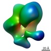 5287MC  5288C M: map data used to model this data C: citing same article ( |
|---|---|
| Similar structure data |

-Links
 PDBj
PDBj


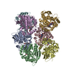
- Assembly
| Deposited unit | 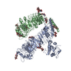
|
|---|---|
| 1 |
|
-Components
| #1: Protein | Mass: 96656.367 Da / Num. of mol.: 2 / Fragment: mature glycoprotein (UNP residues 23-858) Source method: isolated from a genetically manipulated source Source: (gene. exp.) Homo sapiens (human) / Gene: TIL3, TLR5 / Plasmid: pAcGP67-A / Production host:   Spodoptera frugiperda (fall armyworm) / Strain (production host): SF9 / References: UniProt: O60602 Spodoptera frugiperda (fall armyworm) / Strain (production host): SF9 / References: UniProt: O60602#2: Polysaccharide | alpha-L-fucopyranose-(1-6)-2-acetamido-2-deoxy-beta-D-glucopyranose Source method: isolated from a genetically manipulated source Has protein modification | Y | |
|---|
-Experimental details
-Experiment
| Experiment | Method: ELECTRON MICROSCOPY |
|---|---|
| EM experiment | Aggregation state: PARTICLE / 3D reconstruction method: single particle reconstruction |
- Sample preparation
| Component | Name: Full-length Toll-like receptor 5 / Type: COMPLEX / Details: Asymmetric homodimer. The sample was monodisperse. |
|---|---|
| Molecular weight | Value: 0.2 MDa / Experimental value: YES |
| Buffer solution | Name: 10 mM TEA pH 7.5, 0.1 M NaCl, followed by 1% uranyl-formate stain pH: 7.5 Details: 10 mM TEA pH 7.5, 0.1 M NaCl, followed by 1% uranyl-formate stain |
| Specimen | Conc.: 0.005 mg/ml / Embedding applied: NO / Shadowing applied: NO / Staining applied: YES / Vitrification applied: YES / Details: 10 mM TEA, pH 7.5, 0.15 M NaCl, |
| EM staining | Type: NEGATIVE / Material: Uranyl Formate |
| Specimen support | Details: Thin-carbon-covered holey carbon copper grid |
- Electron microscopy imaging
| Microscopy | Model: FEI TECNAI 12 / Date: Aug 1, 2010 |
|---|---|
| Electron gun | Electron source: LAB6 / Accelerating voltage: 120 kV / Illumination mode: FLOOD BEAM |
| Electron lens | Mode: BRIGHT FIELD / Nominal magnification: 52000 X / Calibrated magnification: 52000 X / Nominal defocus max: 1200 nm / Nominal defocus min: 800 nm / Cs: 2.2 mm / Camera length: 0 mm |
| Specimen holder | Specimen holder model: SIDE ENTRY, EUCENTRIC / Specimen holder type: Eucentric / Temperature: 298 K |
| Image recording | Film or detector model: GATAN ULTRASCAN 4000 (4k x 4k) |
| Radiation | Protocol: SINGLE WAVELENGTH / Monochromatic (M) / Laue (L): M / Scattering type: x-ray |
| Radiation wavelength | Relative weight: 1 |
- Processing
| EM software |
| |||||||||||||||
|---|---|---|---|---|---|---|---|---|---|---|---|---|---|---|---|---|
| Symmetry | Point symmetry: C1 (asymmetric) | |||||||||||||||
| 3D reconstruction | Method: Random conical tilt followed by projection matching refinement Resolution: 26 Å / Resolution method: FSC 0.5 CUT-OFF / Num. of particles: 4241 Details: Final maps were calculated from all the particles by back-projection reconstruction. Num. of class averages: 100 / Symmetry type: POINT | |||||||||||||||
| Atomic model building | Protocol: RIGID BODY FIT / Space: REAL / Target criteria: Energy minimization Details: REFINEMENT PROTOCOL--Rigid body and positional refinement DETAILS--Initial local fitting was done using Chimera and then the energy of the model was minimized with MODELLER and REFMAC. | |||||||||||||||
| Refinement step | Cycle: LAST
|
