 Movie
Movie Controller

[English] 日本語
 Yorodumi
- PDB-3izz: Models for ribosome components that are nearest neighbors to the ... -
+ Open data

- Basic information
| Entry | Database: PDB / ID: 3izz | ||||||
|---|---|---|---|---|---|---|---|
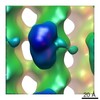
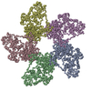
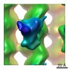
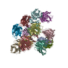
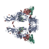
| Title | Models for ribosome components that are nearest neighbors to the bovine mitochondrial initiation factor2 bound to the E. Coli ribosome | ||||||
 Components Components |
| ||||||
 Keywords Keywords | RNA / RIBOSOMAL PROTEIN / 70S | ||||||
| Function / homology |  Function and homology information Function and homology informationlarge ribosomal subunit rRNA binding / cytosolic large ribosomal subunit / cytoplasmic translation / structural constituent of ribosome / cytosol / cytoplasm Similarity search - Function | ||||||
| Biological species |  | ||||||
| Method | ELECTRON MICROSCOPY / single particle reconstruction / cryo EM / Resolution: 10.8 Å | ||||||
 Authors Authors | Yassin, A.S. / Haque, E. / Datta, P.P. / Elmore, K. / Banavali, N.K. / Spremulli, L.L. / Agrawal, R.K. | ||||||
 Citation Citation | Journal: Proc Natl Acad Sci U S A / Year: 2011 Title: Insertion domain within mammalian mitochondrial translation initiation factor 2 serves the role of eubacterial initiation factor 1. Authors: Aymen S Yassin / Md Emdadul Haque / Partha P Datta / Kevin Elmore / Nilesh K Banavali / Linda L Spremulli / Rajendra K Agrawal /  Abstract: Mitochondria have their own translational machineries for the synthesis of thirteen polypeptide chains that are components of the complexes that participate in the process of oxidative ...Mitochondria have their own translational machineries for the synthesis of thirteen polypeptide chains that are components of the complexes that participate in the process of oxidative phosphorylation (or ATP generation). Translation initiation in mammalian mitochondria requires two initiation factors, IF2(mt) and IF3(mt), instead of the three that are present in eubacteria. The mammalian IF2(mt) possesses a unique 37 amino acid insertion domain, which is known to be important for the formation of the translation initiation complex. We have obtained a three-dimensional cryoelectron microscopic map of the mammalian IF2(mt) in complex with initiator fMet-tRNA(iMet) and the eubacterial ribosome. We find that the 37 amino acid insertion domain interacts with the same binding site on the ribosome that would be occupied by the eubacterial initiation factor IF1, which is absent in mitochondria. Our finding suggests that the insertion domain of IF2(mt) mimics the function of eubacterial IF1, by blocking the ribosomal aminoacyl-tRNA binding site (A site) at the initiation step. | ||||||
| History |
|
- Structure visualization
| Movie |
Movie viewer |
|---|---|
| Structure viewer | Molecule: MolmilJmol/JSmol |
- Downloads & links
-Download
| PDBx/mmCIF format | 3izz.cif.gz | 221.8 KB | Display | PDBx/mmCIF format |
|---|---|---|---|---|
| PDB format | pdb3izz.ent.gz | 165.1 KB | Display | PDB format |
| PDBx/mmJSON format | 3izz.json.gz | Tree view | PDBx/mmJSON format | |
| Others |  Other downloads Other downloads |
-Validation report
| Arichive directory | https://data.pdbj.org/pub/pdb/validation_reports/iz/3izzftp://data.pdbj.org/pub/pdb/validation_reports/iz/3izz | HTTPS FTP |
|---|
-Related structure data
| Related structure data |  1854MC  1855C  3izyC M: map data used to model this data C: citing same article ( |
|---|---|
| Similar structure data |


-Links
 PDBj
PDBj


- Assembly
| Deposited unit | 
|
|---|---|
| 1 |
|
-Components
-RNA chain , 4 types, 4 molecules ADEB
| #1: RNA chain | Mass: 19027.375 Da / Num. of mol.: 1 / Source method: isolated from a natural source / Source: (natural) |
|---|---|
| #2: RNA chain | Mass: 19467.643 Da / Num. of mol.: 1 / Source method: isolated from a natural source / Source: (natural) |
| #3: RNA chain | Mass: 32453.391 Da / Num. of mol.: 1 / Source method: isolated from a natural source / Source: (natural) |
| #5: RNA chain | Mass: 38051.535 Da / Num. of mol.: 1 / Source method: isolated from a natural source / Source: (natural) |
-Protein , 2 types, 2 molecules FG
| #4: Protein | Mass: 13804.311 Da / Num. of mol.: 1 / Source method: isolated from a natural source / Source: (natural) |
|---|---|
| #6: Protein | Mass: 13320.714 Da / Num. of mol.: 1 / Source method: isolated from a natural source / Source: (natural) |
-Details
| Sequence details | CHAIN A IS MADE UP OF THREE HELICES (5,14, AND 15) OF SMALL SUBUNIT RNA AND CHAIN B IS COMPOSED OF ...CHAIN A IS MADE UP OF THREE HELICES (5,14, AND 15) OF SMALL SUBUNIT RNA AND CHAIN B IS COMPOSED OF HELICES 69, 71, 89, 92, AND 95 OF LARGE SUBUNIT RIBOSOME. |
|---|
-Experimental details
-Experiment
| Experiment | Method: ELECTRON MICROSCOPY |
|---|---|
| EM experiment | Aggregation state: PARTICLE / 3D reconstruction method: single particle reconstruction |
- Sample preparation
| Component | Name: E. coli 70S / Type: RIBOSOME |
|---|---|
| Buffer solution | pH: 7.6 |
| Specimen | Embedding applied: NO / Shadowing applied: NO / Staining applied: NO / Vitrification applied: YES |
| Vitrification | Instrument: FEI VITROBOT MARK I / Cryogen name: ETHANE / Details: Vitrobot |
- Electron microscopy imaging
| Experimental equipment |  Model: Tecnai F20 / Image courtesy: FEI Company |
|---|---|
| Microscopy | Model: FEI TECNAI F20 |
| Electron gun | Electron source:  FIELD EMISSION GUN / Accelerating voltage: 200 kV / Illumination mode: FLOOD BEAM FIELD EMISSION GUN / Accelerating voltage: 200 kV / Illumination mode: FLOOD BEAM |
| Electron lens | Mode: BRIGHT FIELD / Nominal magnification: 50000 X / Calibrated magnification: 50760 X |
| Image recording | Film or detector model: KODAK SO-163 FILM |
- Processing
| EM software | Name: SPIDER / Category: 3D reconstruction | ||||||||||||
|---|---|---|---|---|---|---|---|---|---|---|---|---|---|
| CTF correction | Details: Every micrograph | ||||||||||||
| Symmetry | Point symmetry: C1 (asymmetric) | ||||||||||||
| 3D reconstruction | Method: Back Projection / Resolution: 10.8 Å / Num. of particles: 121742 / Actual pixel size: 2.76 Å / Symmetry type: POINT | ||||||||||||
| Refinement step | Cycle: LAST
|
