 Movie
Movie Controller


+ Open data

- Basic information
| Entry | Database: PDB / ID: 3izp | ||||||
|---|---|---|---|---|---|---|---|
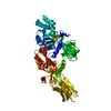
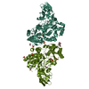
| Title | Conformation of EF-G during translocation | ||||||
 Components Components | Elongation factor G | ||||||
 Keywords Keywords | RIBOSOMAL PROTEIN / electron microscopy / flexible fitting / GTP hydrolysis / hybrid state / inter-subunit rotation / ribosome / translation / tRNA | ||||||
| Function / homology |  Function and homology information Function and homology informationribosome disassembly / translational elongation / translation elongation factor activity / GDP binding / ribosome binding / GTPase activity / GTP binding / magnesium ion binding / cytoplasm Similarity search - Function | ||||||
| Biological species |   Thermus thermophilus (bacteria) Thermus thermophilus (bacteria) | ||||||
| Method | ELECTRON MICROSCOPY / single particle reconstruction / cryo EM | ||||||
 Authors Authors | Li, W. / Trabuco, L.G. / Schulten, K. / Frank, J. | ||||||
 Citation Citation | Journal: Proteins / Year: 2011 Title: Molecular dynamics of EF-G during translocation. Authors: Wen Li / Leonardo G Trabuco / Klaus Schulten / Joachim Frank /  Abstract: Elongation factor G (EF-G) plays a crucial role in two stages of mRNA-(tRNA)(2) translocation. First, EF-G•GTP enters the pre-translocational ribosome in its intersubunit-rotated state, with tRNAs ...Elongation factor G (EF-G) plays a crucial role in two stages of mRNA-(tRNA)(2) translocation. First, EF-G•GTP enters the pre-translocational ribosome in its intersubunit-rotated state, with tRNAs in their hybrid (P/E and A/P) positions. Second, a conformational change in EF-G's Domain IV induced by GTP hydrolysis disengages the mRNA-anticodon stem-loops of the tRNAs from the decoding center to advance relative to the small subunit when the ribosome undergoes a backward inter-subunit rotation. These events take place as EF-G undergoes a series of large conformational changes as visualized by cryo-EM and X-ray studies. The number and variety of these structures leave open many questions on how these different configurations form during the dynamic translocation process. To understand the molecular mechanism of translocation, we examined the molecular motions of EF-G in solution by means of molecular dynamics simulations. Our results show: (1) rotations of the super-domain formed by Domains III-V with respect to the super-domain formed by I-II, and rotations of Domain IV with respect to Domain III; (2) flexible conformations of both 503- and 575-loops; (3) large conformational variability in the bound form caused by the interaction between Domain V and the GTPase-associated center; (4) after GTP hydrolysis, the Switch I region seems to be instrumental for effecting the conformational change at the end of Domain IV implicated in the disengagement of the codon-anticodon helix from the decoding center. | ||||||
| History |
|
- Structure visualization
| Movie |
Movie viewer |
|---|---|
| Structure viewer | Molecule: MolmilJmol/JSmol |
- Downloads & links
-Download
| PDBx/mmCIF format | 3izp.cif.gz | 128.8 KB | Display | PDBx/mmCIF format |
|---|---|---|---|---|
| PDB format | pdb3izp.ent.gz | 96.4 KB | Display | PDB format |
| PDBx/mmJSON format | 3izp.json.gz | Tree view | PDBx/mmJSON format | |
| Others |  Other downloads Other downloads |
-Validation report
| Arichive directory | https://data.pdbj.org/pub/pdb/validation_reports/iz/3izpftp://data.pdbj.org/pub/pdb/validation_reports/iz/3izp | HTTPS FTP |
|---|
-Related structure data
| Related structure data |  1363F F: fitted*YM |
|---|---|
| Similar structure data |


-Links
 PDBj
PDBj


- Assembly
| Deposited unit | 
|
|---|---|
| 1 |
|
-Components
| #1: Protein | Mass: 76595.664 Da / Num. of mol.: 1 / Source method: isolated from a natural source / Source: (natural) Thermus thermophilus (bacteria) / References: UniProt: P13551 |
|---|
-Experimental details
-Experiment
| Experiment | Method: ELECTRON MICROSCOPY |
|---|---|
| EM experiment | Aggregation state: PARTICLE / 3D reconstruction method: single particle reconstruction |
- Sample preparation
| Component | Name: 70S ribosome / Type: RIBOSOME |
|---|---|
| Buffer solution | pH: 7.5 Details: 5 mM potassium phosphate, 5 mM magnesium acetate, 5 mM ammonium chloride, 95 mM potassium chloride, 0.5 mM calcium chloride, 8 mM putrescine, 1 mM spermidine, and 1 mM dithioerythritol |
| Specimen | Embedding applied: NO / Shadowing applied: NO / Staining applied: NO / Vitrification applied: YES |
| Vitrification | Cryogen name: ETHANE / Temp: 90 K / Method: two-face blotting for 1 second |
- Electron microscopy imaging
| Experimental equipment |  Model: Tecnai F20 / Image courtesy: FEI Company |
|---|---|
| Microscopy | Model: FEI TECNAI F20 |
| Electron gun | Electron source:  FIELD EMISSION GUN / Accelerating voltage: 200 kV / Illumination mode: FLOOD BEAM FIELD EMISSION GUN / Accelerating voltage: 200 kV / Illumination mode: FLOOD BEAM |
| Electron lens | Mode: BRIGHT FIELD / Nominal magnification: 50000 X / Calibrated magnification: 49650 X / Nominal defocus max: 4000 nm / Nominal defocus min: 2000 nm / Cs: 2 mm |
| Specimen holder | Specimen holder model: GATAN LIQUID NITROGEN / Specimen holder type: single tilt cryoholder / Tilt angle max: 0 ° / Tilt angle min: 0 ° |
| Image recording | Electron dose: 15 e/Å2 / Film or detector model: KODAK SO-163 FILM |
| Radiation | Protocol: SINGLE WAVELENGTH / Monochromatic (M) / Laue (L): M |
| Radiation wavelength | Relative weight: 1 |
- Processing
| EM software | Name: MDFF / Category: model fitting | ||||||||||||
|---|---|---|---|---|---|---|---|---|---|---|---|---|---|
| Symmetry | Point symmetry: C1 (asymmetric) | ||||||||||||
| 3D reconstruction | Symmetry type: POINT | ||||||||||||
| Atomic model building | Protocol: FLEXIBLE FIT / Space: REAL Details: REFINEMENT PROTOCOL--Molecular Dynamics Flexible Fitting | ||||||||||||
| Atomic model building | PDB-ID: 1FNM Accession code: 1FNM / Source name: PDB / Type: experimental model | ||||||||||||
| Refinement step | Cycle: LAST
|
