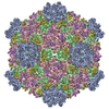
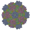
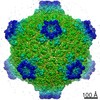
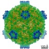
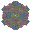
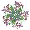
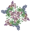
ジャーナル: Proc Natl Acad Sci U S A / 年: 2011 タイトル: Atomic model of a cypovirus built from cryo-EM structure provides insight into the mechanism of mRNA capping. 著者: Lingpeng Cheng / Jingchen Sun / Kai Zhang / Zongjun Mou / Xiaoxing Huang / Gang Ji / Fei Sun / Jingqiang Zhang / Ping Zhu / 要旨: The cytoplasmic polyhedrosis virus (CPV) from the family Reoviridae belongs to a subgroup of "turreted" reoviruses, in which the mRNA capping activity occurs in a pentameric turret. We report a full ...The cytoplasmic polyhedrosis virus (CPV) from the family Reoviridae belongs to a subgroup of "turreted" reoviruses, in which the mRNA capping activity occurs in a pentameric turret. We report a full atomic model of CPV built from a 3D density map obtained using cryoelectron microscopy. The image data for the 3D reconstruction were acquired exclusively from a CCD camera. Our structure shows that the enzymatic domains of the pentameric turret of CPV are topologically conserved and that there are five unique channels connecting the guanylyltransferase and methyltransferase regions. This structural organization reveals how the channels guide nascent mRNA sequentially to guanylyltransferase, 7-N-methyltransferase, and 2'-O-methyltransferase in the turret, undergoing the highly coordinated mRNA capping activity. Furthermore, by fitting the deduced amino acid sequence of the protein VP5 to 120 large protrusion proteins on the CPV capsid shell, we confirmed that this protrusion protein is encoded by CPV RNA segment 7.
 ムービー
ムービー コントローラー


 データを開く
 基本情報
基本情報 要素
要素 キーワード
キーワード 機能・相同性情報
 Bombyx mori cypovirus 1 (ウイルス)
 データ登録者
 引用
引用
 構造の表示
構造の表示 ダウンロードとリンク
 その他のダウンロード


 PDBj
PDBj
 集合体
集合体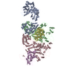

 試料調製
試料調製 電子顕微鏡撮影
 FIELD EMISSION GUN / 加速電圧: 300 kV / 照射モード: OTHER
 解析
解析