 Movie
Movie Controller


+ Open data

- Basic information
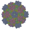
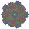
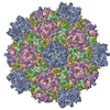
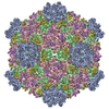
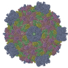
| Entry | Database: PDB / ID: 3jb1 | ||||||
|---|---|---|---|---|---|---|---|
| Title | Atomic model of cytoplasmic polyhedrosis virus with SAM | ||||||
 Components Components |
| ||||||
 Keywords Keywords | VIRUS / viral ATPase / histidine-mediated guanylyl transfer / conformational changes / regulation of transcription | ||||||
| Function / homology |  Function and homology information Function and homology information | ||||||
| Biological species |   Bombyx mori cypovirus 1 Bombyx mori cypovirus 1 | ||||||
| Method | ELECTRON MICROSCOPY / single particle reconstruction / cryo EM / Resolution: 3.1 Å | ||||||
 Authors Authors | Yu, X.K. / Jiang, J.S. / Sun, J.C. / Zhou, Z.H. | ||||||
 Citation Citation | Journal: Elife / Year: 2015 Title: A putative ATPase mediates RNA transcription and capping in a dsRNA virus. Authors: Xuekui Yu / Jiansen Jiang / Jingchen Sun / Z Hong Zhou /  Abstract: mRNA transcription in dsRNA viruses is a highly regulated process but the mechanism of this regulation is not known. Here, by nucleoside triphosphatase (NTPase) assay and comparisons of six high- ...mRNA transcription in dsRNA viruses is a highly regulated process but the mechanism of this regulation is not known. Here, by nucleoside triphosphatase (NTPase) assay and comparisons of six high-resolution (2.9-3.1 Å) cryo-electron microscopy structures of cytoplasmic polyhedrosis virus with bound ligands, we show that the large sub-domain of the guanylyltransferase (GTase) domain of the turret protein (TP) also has an ATP-binding site and is likely an ATPase. S-adenosyl-L-methionine (SAM) acts as a signal and binds the methylase-2 domain of TP to induce conformational change of the viral capsid, which in turn activates the putative ATPase. ATP binding/hydrolysis leads to an enlarged capsid for efficient mRNA synthesis, an open GTase domain for His217-mediated guanylyl transfer, and an open methylase-1 domain for SAM binding and methyl transfer. Taken together, our data support a role of the putative ATPase in mediating the activation of mRNA transcription and capping within the confines of the virus. | ||||||
| History |
|
- Structure visualization
| Movie |
Movie viewer |
|---|---|
| Structure viewer | Molecule: MolmilJmol/JSmol |
- Downloads & links
-Download
| PDBx/mmCIF format | 3jb1.cif.gz | 822.1 KB | Display | PDBx/mmCIF format |
|---|---|---|---|---|
| PDB format | pdb3jb1.ent.gz | 659.6 KB | Display | PDB format |
| PDBx/mmJSON format | 3jb1.json.gz | Tree view | PDBx/mmJSON format | |
| Others |  Other downloads Other downloads |
-Validation report
| Arichive directory | https://data.pdbj.org/pub/pdb/validation_reports/jb/3jb1ftp://data.pdbj.org/pub/pdb/validation_reports/jb/3jb1 | HTTPS FTP |
|---|
-Related structure data
| Related structure data | 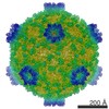 6375MC  6371C 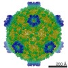 6374C 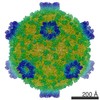 6376C 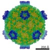 6377C 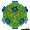 6378C  3jayC  3jazC  3jb0C  3jb2C  3jb3C M: map data used to model this data C: citing same article ( |
|---|---|
| Similar structure data |
-Links
 PDBj
PDBj

- Assembly
| Deposited unit | 
|
|---|---|
| 1 | x 60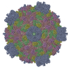
|
| 2 |
|
| 3 | x 5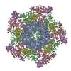
|
| 4 | x 6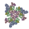
|
| 5 | 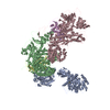
|
| Symmetry | Point symmetry: (Schoenflies symbol: I (icosahedral)) |
-Components
| #1: Protein | Mass: 120145.797 Da / Num. of mol.: 1 / Source method: isolated from a natural source / Source: (natural) Bombyx mori cypovirus 1 / References: UniProt: Q914N6 | ||||
|---|---|---|---|---|---|
| #2: Protein | Mass: 148560.859 Da / Num. of mol.: 2 / Source method: isolated from a natural source / Source: (natural) Bombyx mori cypovirus 1 / References: UniProt: Q6TS43#3: Protein | Mass: 49906.176 Da / Num. of mol.: 2 / Source method: isolated from a natural source / Source: (natural) Bombyx mori cypovirus 1 / References: UniProt: C6K2M8#4: Chemical | ChemComp-SAM / |   Mass: 398.437 Da / Num. of mol.: 1 / Source method: obtained synthetically / Formula: C15H22N6O5S Mass: 398.437 Da / Num. of mol.: 1 / Source method: obtained synthetically / Formula: C15H22N6O5S |
-Experimental details
-Experiment
| Experiment | Method: ELECTRON MICROSCOPY |
|---|---|
| EM experiment | Aggregation state: PARTICLE / 3D reconstruction method: single particle reconstruction |
- Sample preparation
| Component |
| |||||||||||||||
|---|---|---|---|---|---|---|---|---|---|---|---|---|---|---|---|---|
| Details of virus | Empty: NO / Enveloped: NO / Host category: INVERTEBRATES / Isolate: SPECIES / Type: VIRION | |||||||||||||||
| Natural host | Organism: Bombyx mori | |||||||||||||||
| Specimen | Embedding applied: NO / Shadowing applied: NO / Staining applied: NO / Vitrification applied: YES | |||||||||||||||
| Vitrification | Instrument: FEI VITROBOT MARK II / Cryogen name: ETHANE / Humidity: 100 % / Details: Plunged into liquid ethane (FEI VITROBOT MARK II) |
- Electron microscopy imaging
| Experimental equipment |  Model: Titan Krios / Image courtesy: FEI Company |
|---|---|
| Microscopy | Model: FEI TITAN KRIOS / Date: May 8, 2012 |
| Electron gun | Electron source:  FIELD EMISSION GUN / Accelerating voltage: 300 kV / Illumination mode: FLOOD BEAM FIELD EMISSION GUN / Accelerating voltage: 300 kV / Illumination mode: FLOOD BEAM |
| Electron lens | Mode: BRIGHT FIELD / Nominal magnification: 59000 X / Calibrated magnification: 60535 X / Cs: 2.75 mm Astigmatism: Objective lens astigmatism was corrected at 135,000 times magnification. |
| Specimen holder | Specimen holder model: FEI TITAN KRIOS AUTOGRID HOLDER |
| Image recording | Electron dose: 25 e/Å2 / Film or detector model: KODAK SO-163 FILM |
| Radiation | Protocol: SINGLE WAVELENGTH / Monochromatic (M) / Laue (L): M / Scattering type: x-ray |
| Radiation wavelength | Relative weight: 1 |
- Processing
| EM software | Name: IMIRS / Category: 3D reconstruction | ||||||||||||||||||||||||||||||||||||||||||||||||||||||||||||||||||||||||||||||
|---|---|---|---|---|---|---|---|---|---|---|---|---|---|---|---|---|---|---|---|---|---|---|---|---|---|---|---|---|---|---|---|---|---|---|---|---|---|---|---|---|---|---|---|---|---|---|---|---|---|---|---|---|---|---|---|---|---|---|---|---|---|---|---|---|---|---|---|---|---|---|---|---|---|---|---|---|---|---|---|
| CTF correction | Details: Each particle | ||||||||||||||||||||||||||||||||||||||||||||||||||||||||||||||||||||||||||||||
| Symmetry | Point symmetry: I (icosahedral) | ||||||||||||||||||||||||||||||||||||||||||||||||||||||||||||||||||||||||||||||
| 3D reconstruction | Method: Cross-common lines / Resolution: 3.1 Å / Resolution method: FSC 0.143 CUT-OFF / Num. of particles: 44908 / Nominal pixel size: 1.104 Å / Actual pixel size: 1.104 Å / Details: (Single particle--Applied symmetry: I) / Symmetry type: POINT | ||||||||||||||||||||||||||||||||||||||||||||||||||||||||||||||||||||||||||||||
| Refinement | Resolution: 3.1→39.032 Å / SU ML: 0.57 / σ(F): 2 / Phase error: 27.67 / Stereochemistry target values: MLHL
| ||||||||||||||||||||||||||||||||||||||||||||||||||||||||||||||||||||||||||||||
| Solvent computation | Shrinkage radii: 0.9 Å / VDW probe radii: 1.11 Å / Solvent model: FLAT BULK SOLVENT MODEL | ||||||||||||||||||||||||||||||||||||||||||||||||||||||||||||||||||||||||||||||
| Displacement parameters | Biso max: 530.97 Å2 / Biso mean: 204.9893 Å2 / Biso min: 126.4 Å2 | ||||||||||||||||||||||||||||||||||||||||||||||||||||||||||||||||||||||||||||||
| Refinement step | Cycle: LAST / Resolution: 3.1→39.032 Å
| ||||||||||||||||||||||||||||||||||||||||||||||||||||||||||||||||||||||||||||||
| Refine LS restraints |
| ||||||||||||||||||||||||||||||||||||||||||||||||||||||||||||||||||||||||||||||
| LS refinement shell | Refine-ID: ELECTRON MICROSCOPY / Total num. of bins used: 12 / % reflection obs: 100 %
| ||||||||||||||||||||||||||||||||||||||||||||||||||||||||||||||||||||||||||||||
| Refinement TLS params. | Method: refined / Origin x: -42.333 Å / Origin y: -68.136 Å / Origin z: 259.7022 Å
| ||||||||||||||||||||||||||||||||||||||||||||||||||||||||||||||||||||||||||||||
| Refinement TLS group |
|
