 Movie
Movie Controller


+ Open data

- Basic information
| Entry | Database: PDB / ID: 3iz2 | ||||||
|---|---|---|---|---|---|---|---|
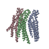
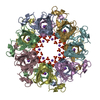
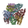
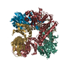
| Title | C-alpha model fitted into the EM structure of Cx26M34Adel2-7 | ||||||
 Components Components | Gap junction beta-2 protein | ||||||
 Keywords Keywords | MEMBRANE PROTEIN / Gap Junction channel | ||||||
| Function / homology |  Function and homology information Function and homology informationTransport of connexons to the plasma membrane / gap junction channel activity involved in cell communication by electrical coupling / gap junction-mediated intercellular transport / Oligomerization of connexins into connexons / Transport of connexins along the secretory pathway / gap junction assembly / connexin complex / gap junction / Gap junction assembly / gap junction channel activity ...Transport of connexons to the plasma membrane / gap junction channel activity involved in cell communication by electrical coupling / gap junction-mediated intercellular transport / Oligomerization of connexins into connexons / Transport of connexins along the secretory pathway / gap junction assembly / connexin complex / gap junction / Gap junction assembly / gap junction channel activity / endoplasmic reticulum-Golgi intermediate compartment / sensory perception of sound / transmembrane transport / cell-cell signaling / calcium ion binding / identical protein binding / plasma membrane Similarity search - Function | ||||||
| Biological species |  Homo sapiens (human) Homo sapiens (human) | ||||||
| Method | ELECTRON CRYSTALLOGRAPHY / electron crystallography / cryo EM / Resolution: 10 Å | ||||||
 Authors Authors | Oshima, A. / Tani, K. / Toloue, M.M. / Hiroaki, Y. / Smock, A. / Inukai, S. / Cone, A. / Nicholson, B.J. / Sosinsky, G.E. / Fujiyoshi, Y. | ||||||
 Citation Citation | Journal: J Mol Biol / Year: 2011 Title: Asymmetric configurations and N-terminal rearrangements in connexin26 gap junction channels. Authors: Atsunori Oshima / Kazutoshi Tani / Masoud M Toloue / Yoko Hiroaki / Amy Smock / Sayaka Inukai / Angela Cone / Bruce J Nicholson / Gina E Sosinsky / Yoshinori Fujiyoshi /  Abstract: Gap junction channels are unique in that they possess multiple mechanisms for channel closure, several of which involve the N terminus as a key component in gating, and possibly assembly. Here, we ...Gap junction channels are unique in that they possess multiple mechanisms for channel closure, several of which involve the N terminus as a key component in gating, and possibly assembly. Here, we present electron crystallographic structures of a mutant human connexin26 (Cx26M34A) and an N-terminal deletion of this mutant (Cx26M34Adel2-7) at 6-Å and 10-Å resolutions, respectively. The three-dimensional map of Cx26M34A was improved by data from 60° tilt images and revealed a breakdown of the hexagonal symmetry in a connexin hemichannel, particularly in the cytoplasmic domain regions at the ends of the transmembrane helices. The Cx26M34A structure contained an asymmetric density in the channel vestibule ("plug") that was decreased in the Cx26M34Adel2-7 structure, indicating that the N terminus significantly contributes to form this plug feature. Functional analysis of the Cx26M34A channels revealed that these channels are predominantly closed, with the residual electrical conductance showing normal voltage gating. N-terminal deletion mutants with and without the M34A mutation showed no electrical activity in paired Xenopus oocytes and significantly decreased dye permeability in HeLa cells. Comparing this closed structure with the recently published X-ray structure of wild-type Cx26, which is proposed to be in an open state, revealed a radial outward shift in the transmembrane helices in the closed state, presumably to accommodate the N-terminal plug occluding the pore. Because both Cx26del2-7 and Cx26M34Adel2-7 channels are closed, the N terminus appears to have a prominent role in stabilizing the open configuration. | ||||||
| History |
|
- Structure visualization
| Movie |
Movie viewer |
|---|---|
| Structure viewer | Molecule: MolmilJmol/JSmol |
- Downloads & links
-Download
| PDBx/mmCIF format | 3iz2.cif.gz | 31.4 KB | Display | PDBx/mmCIF format |
|---|---|---|---|---|
| PDB format | pdb3iz2.ent.gz | 16.4 KB | Display | PDB format |
| PDBx/mmJSON format | 3iz2.json.gz | Tree view | PDBx/mmJSON format | |
| Others |  Other downloads Other downloads |
-Validation report
| Arichive directory | https://data.pdbj.org/pub/pdb/validation_reports/iz/3iz2ftp://data.pdbj.org/pub/pdb/validation_reports/iz/3iz2 | HTTPS FTP |
|---|
-Related structure data
| Related structure data |  1749MC  1748C  3iz1C M: map data used to model this data C: citing same article ( |
|---|---|
| Similar structure data |


-Links
 PDBj
PDBj- Assembly
| Deposited unit | 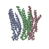
| ||||||||
|---|---|---|---|---|---|---|---|---|---|
| 1 | 
| ||||||||
| Unit cell |
|
-Components
| #1: Protein | Mass: 26985.943 Da / Num. of mol.: 3 / Mutation: M34A Source method: isolated from a genetically manipulated source Source: (gene. exp.) Homo sapiens (human) / Gene: GJB2 / Plasmid: pBlueBac4.5 / Production host:   Spodoptera frugiperda (fall armyworm) / References: UniProt: P29033 Spodoptera frugiperda (fall armyworm) / References: UniProt: P29033 |
|---|
-Experimental details
-Experiment
| Experiment | Method: ELECTRON CRYSTALLOGRAPHY |
|---|---|
| EM experiment | Aggregation state: 2D ARRAY / 3D reconstruction method: electron crystallography |
- Sample preparation
| Component | Name: connexin26 gap junction channels / Type: COMPLEX |
|---|---|
| Buffer solution | Name: 10mM MES / pH: 5.8 Details: 10% trehalose (Sigma, St. Louis, MO), 10 mM MES (pH 5.8), 300 mM NaCl, 50 mM MgCl2, 5 mM CaCl2, 2 mM dithiothreitol, 100 microM carbenoxolone, 0.005% NaN3, and 1% glycerol10mM MES |
| Specimen | Conc.: 2 mg/ml / Embedding applied: YES / Shadowing applied: NO / Staining applied: NO / Vitrification applied: YES |
| Specimen support | Grid material: MOLYBDENUM |
| EM embedding | Details: 10% trehalose / Material: trehalose |
| Vitrification | Instrument: REICHERT-JUNG PLUNGER / Cryogen name: NITROGEN / Temp: 93 K Method: The grids were blotted with filter paper and fast frozen into liquid nitrogen |
-Data collection
| Microscopy | Model: JEOL KYOTO-3000SFF / Date: Nov 9, 2007 |
|---|---|
| Electron gun | Electron source:  FIELD EMISSION GUN / Accelerating voltage: 300 kV / Illumination mode: FLOOD BEAM FIELD EMISSION GUN / Accelerating voltage: 300 kV / Illumination mode: FLOOD BEAM |
| Electron lens | Mode: BRIGHT FIELD / Nominal magnification: 40000 X / Calibrated magnification: 39000 X / Nominal defocus max: 2214 nm / Nominal defocus min: 544 nm / Cs: 1.6 mm |
| Specimen holder | Temperature: 4.2 K / Tilt angle max: 45 ° / Tilt angle min: 0 ° |
| Image recording | Electron dose: 25 e/Å2 / Film or detector model: KODAK SO-163 FILM |
| Radiation | Protocol: SINGLE WAVELENGTH / Monochromatic (M) / Laue (L): M / Scattering type: electron |
| Radiation wavelength | Relative weight: 1 |
- Processing
| EM software |
| ||||||||||||
|---|---|---|---|---|---|---|---|---|---|---|---|---|---|
| CTF correction | Details: Each image | ||||||||||||
| 3D reconstruction | Method: Lattice line fitting / Resolution: 10 Å / Magnification calibration: carbon grating / Symmetry type: 2D CRYSTAL | ||||||||||||
| Atomic model building | Protocol: RIGID BODY FIT / Space: REAL / Target criteria: Cross-correlation coefficient Details: REFINEMENT PROTOCOL--Rigid body DETAILS--Initial local fitting was done using O, and Situs was used for flexible fitting. | ||||||||||||
| Atomic model building | PDB-ID: 2ZW3 Accession code: 2ZW3 / Source name: PDB / Type: experimental model | ||||||||||||
| Refinement step | Cycle: LAST
|

