 Movie
Movie Controller

[English] 日本語
 Yorodumi
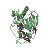
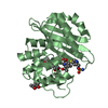
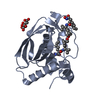
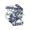
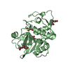
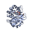
- PDB-3ixm: Structure of the Gly74Cys mutant of arylmalonate decarboxylase in... -
+ Open data

- Basic information
| Entry | Database: PDB / ID: 3ixm | ||||||
|---|---|---|---|---|---|---|---|
| Title | Structure of the Gly74Cys mutant of arylmalonate decarboxylase in the sulfate ion associated form | ||||||
 Components Components | Arylmalonate decarboxylase | ||||||
 Keywords Keywords | LYASE / enantioselective racemase / Decarboxylase | ||||||
| Function / homology |  Function and homology information Function and homology information | ||||||
| Biological species |  Bordetella bronchiseptica (bacteria) Bordetella bronchiseptica (bacteria) | ||||||
| Method |  X-RAY DIFFRACTION / SYNCHROTRON / MOLECULAR REPLACEMENT / Resolution: 1.9 Å X-RAY DIFFRACTION / SYNCHROTRON / MOLECULAR REPLACEMENT / Resolution: 1.9 Å | ||||||
 Authors Authors | Nakasako, M. / Obata, R. | ||||||
 Citation Citation | Journal: Biochemistry / Year: 2010 Title: Structural Basis for Inverting the Enantioselectivity of Arylmalonate Decarboxylase Revealed by the Structural Analysis of the Gly74Cys/Cys188Ser Mutant in the Liganded Form Authors: Obata, R. / Nakasako, M. #1: Journal: Acta Crystallogr.,Sect.F / Year: 2008 Title: Crystallization and preliminary X-ray diffraction experiments of arylmalonate decarboxylase from Alcaligenes bronchisepticus Authors: Nakasako, M. / Obata, R. / Okubo, R. / Nakayama, S. / Miyamoto, K. / Ohta, H. | ||||||
| History |
|
- Structure visualization
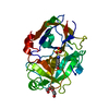
| Structure viewer | Molecule: MolmilJmol/JSmol |
|---|
- Downloads & links
-Download
| PDBx/mmCIF format | 3ixm.cif.gz | 58.9 KB | Display | PDBx/mmCIF format |
|---|---|---|---|---|
| PDB format | pdb3ixm.ent.gz | 41.9 KB | Display | PDB format |
| PDBx/mmJSON format | 3ixm.json.gz | Tree view | PDBx/mmJSON format | |
| Others |  Other downloads Other downloads |
-Validation report
| Arichive directory | https://data.pdbj.org/pub/pdb/validation_reports/ix/3ixmftp://data.pdbj.org/pub/pdb/validation_reports/ix/3ixm | HTTPS FTP |
|---|
-Related structure data
| Related structure data |  3dtvSC  3ixlC C: citing same article ( S: Starting model for refinement |
|---|---|
| Similar structure data |


-Links
 PDBj
PDBj
- Assembly
| Deposited unit | 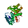
| ||||||||
|---|---|---|---|---|---|---|---|---|---|
| 1 |
| ||||||||
| Unit cell |
|
-Components
| #1: Protein | Mass: 24801.449 Da / Num. of mol.: 1 / Mutation: G74C Source method: isolated from a genetically manipulated source Source: (gene. exp.) Bordetella bronchiseptica (bacteria) / Strain: KU1201 / Plasmid: pAMD101 / Production host: | ||
|---|---|---|---|
| #2: Chemical |   Mass: 96.063 Da / Num. of mol.: 2 / Source method: obtained synthetically / Formula: SO4 Mass: 96.063 Da / Num. of mol.: 2 / Source method: obtained synthetically / Formula: SO4#3: Water | ChemComp-HOH / |  Mass: 18.015 Da / Num. of mol.: 161 / Source method: isolated from a natural source / Formula: H2O Mass: 18.015 Da / Num. of mol.: 161 / Source method: isolated from a natural source / Formula: H2O |
-Experimental details
-Experiment
| Experiment | Method: X-RAY DIFFRACTION / Number of used crystals: 1 |
|---|
- Sample preparation
| Crystal | Density Matthews: 2.13 Å3/Da / Density % sol: 42.32 % |
|---|---|
| Crystal grow | Temperature: 293 K / Method: vapor diffusion, hanging drop / pH: 6.2 Details: 1.2M ammonium sulfate, 15%(w/v) glycerol, 0.1M HEPES, pH 6.2, VAPOR DIFFUSION, HANGING DROP, temperature 293K |
-Data collection
| Diffraction | Mean temperature: 100 K |
|---|---|
| Diffraction source | Source: SYNCHROTRON / Site: SPring-8  / Beamline: BL26B1 / Wavelength: 1 Å / Beamline: BL26B1 / Wavelength: 1 Å |
| Detector | Type: MARMOSAIC 225 mm CCD / Detector: CCD / Date: Apr 22, 2008 / Details: mirror |
| Radiation | Monochromator: Si / Protocol: SINGLE WAVELENGTH / Monochromatic (M) / Laue (L): M / Scattering type: x-ray |
| Radiation wavelength | Wavelength: 1 Å / Relative weight: 1 |
| Reflection | Resolution: 1.9→100 Å / Num. obs: 16054 / % possible obs: 94.2 % / Observed criterion σ(F): 1 / Rmerge(I) obs: 0.053 / Num. measured all: 174138 |
| Reflection shell | Resolution: 1.9→1.93 Å / Rmerge(I) obs: 0.129 / % possible all: 100 |
- Processing
| Software |
| ||||||||||||||||||||||||||||||||||||||||||||||||||||||||||||||||||||||||||||||||||||||||||
|---|---|---|---|---|---|---|---|---|---|---|---|---|---|---|---|---|---|---|---|---|---|---|---|---|---|---|---|---|---|---|---|---|---|---|---|---|---|---|---|---|---|---|---|---|---|---|---|---|---|---|---|---|---|---|---|---|---|---|---|---|---|---|---|---|---|---|---|---|---|---|---|---|---|---|---|---|---|---|---|---|---|---|---|---|---|---|---|---|---|---|---|
| Refinement | Method to determine structure: MOLECULAR REPLACEMENT Starting model: PDB ENTRY 3DTV Resolution: 1.9→20 Å / Cor.coef. Fo:Fc: 0.952 / Cor.coef. Fo:Fc free: 0.916 / SU B: 3.484 / SU ML: 0.106 / Cross valid method: THROUGHOUT / ESU R: 0.17 / ESU R Free: 0.165 / Stereochemistry target values: MAXIMUM LIKELIHOOD / Details: HYDROGENS HAVE BEEN ADDED IN THE RIDING POSITIONS
| ||||||||||||||||||||||||||||||||||||||||||||||||||||||||||||||||||||||||||||||||||||||||||
| Solvent computation | Ion probe radii: 0.8 Å / Shrinkage radii: 0.8 Å / VDW probe radii: 1.2 Å / Solvent model: MASK | ||||||||||||||||||||||||||||||||||||||||||||||||||||||||||||||||||||||||||||||||||||||||||
| Displacement parameters | Biso mean: 23.296 Å2
| ||||||||||||||||||||||||||||||||||||||||||||||||||||||||||||||||||||||||||||||||||||||||||
| Refinement step | Cycle: LAST / Resolution: 1.9→20 Å
| ||||||||||||||||||||||||||||||||||||||||||||||||||||||||||||||||||||||||||||||||||||||||||
| Refine LS restraints |
| ||||||||||||||||||||||||||||||||||||||||||||||||||||||||||||||||||||||||||||||||||||||||||
| LS refinement shell | Resolution: 1.902→1.951 Å / Total num. of bins used: 20
|
