 Movie
Movie Controller

[English] 日本語
 Yorodumi
- PDB-3ix9: Crystal structure of Streptococcus pneumoniae dihydrofolate reduc... -
+ Open data

- Basic information
| Entry | Database: PDB / ID: 3ix9 | ||||||
|---|---|---|---|---|---|---|---|
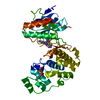
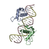
| Title | Crystal structure of Streptococcus pneumoniae dihydrofolate reductase - Sp9 mutant | ||||||
 Components Components | Dihydrofolate reductase | ||||||
 Keywords Keywords | OXIDOREDUCTASE / Central beta sheet surrounded by 4 alpha helices | ||||||
| Function / homology |  Function and homology information Function and homology informationdihydrofolate metabolic process / dihydrofolate reductase / dihydrofolate reductase activity / folic acid metabolic process / tetrahydrofolate biosynthetic process / one-carbon metabolic process / NADP binding / response to antibiotic / cytosol Similarity search - Function | ||||||
| Biological species |   Streptococcus pneumoniae (bacteria) Streptococcus pneumoniae (bacteria) | ||||||
| Method |  X-RAY DIFFRACTION / MOLECULAR REPLACEMENT / Resolution: 1.95 Å X-RAY DIFFRACTION / MOLECULAR REPLACEMENT / Resolution: 1.95 Å | ||||||
 Authors Authors | Yennawar, N.H. | ||||||
 Citation Citation | Journal: Biochemistry / Year: 2010 Title: Kinetic and structural characterization of dihydrofolate reductase from Streptococcus pneumoniae Authors: Lee, J. / Yennawar, N.H. / Gam, J. / Benkovic, S.J. | ||||||
| History |
|
- Structure visualization
| Structure viewer | Molecule: MolmilJmol/JSmol |
|---|
- Downloads & links
-Download
| PDBx/mmCIF format | 3ix9.cif.gz | 93.9 KB | Display | PDBx/mmCIF format |
|---|---|---|---|---|
| PDB format | pdb3ix9.ent.gz | 70.3 KB | Display | PDB format |
| PDBx/mmJSON format | 3ix9.json.gz | Tree view | PDBx/mmJSON format | |
| Others |  Other downloads Other downloads |
-Validation report
| Summary document | 3ix9_validation.pdf.gz | 1.6 MB | Display | wwPDB validaton report |
|---|---|---|---|---|
| Full document | 3ix9_full_validation.pdf.gz | 1.6 MB | Display | |
| Data in XML | 3ix9_validation.xml.gz | 20.3 KB | Display | |
| Data in CIF | 3ix9_validation.cif.gz | 28.2 KB | Display | |
| Arichive directory | https://data.pdbj.org/pub/pdb/validation_reports/ix/3ix9ftp://data.pdbj.org/pub/pdb/validation_reports/ix/3ix9 | HTTPS FTP |
-Related structure data
| Related structure data | 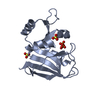 1zdrS S: Starting model for refinement |
|---|---|
| Similar structure data |

-Links
 PDBj
PDBj

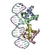
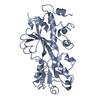
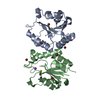
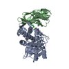
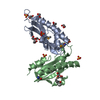
- Assembly
| Deposited unit | 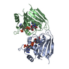
| ||||||||
|---|---|---|---|---|---|---|---|---|---|
| 1 |
| ||||||||
| 2 | 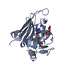
| ||||||||
| 3 | 
| ||||||||
| Unit cell |
| ||||||||
| Components on special symmetry positions |
|
-Components
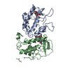
| #1: Protein | Mass: 22230.260 Da / Num. of mol.: 2 / Mutation: E54G, I63T, Q91S, L100V, E133A, A149T Source method: isolated from a genetically manipulated source Source: (gene. exp.) Streptococcus pneumoniae (bacteria) / Gene: dfr / Plasmid: pET28a / Production host: References: UniProt: O85261, UniProt: Q54801*PLUS, dihydrofolate reductase #2: Chemical |   Mass: 745.421 Da / Num. of mol.: 2 / Source method: obtained synthetically / Formula: C21H30N7O17P3 Mass: 745.421 Da / Num. of mol.: 2 / Source method: obtained synthetically / Formula: C21H30N7O17P3#3: Chemical | 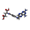  Mass: 454.439 Da / Num. of mol.: 2 / Source method: obtained synthetically / Formula: C20H22N8O5 Mass: 454.439 Da / Num. of mol.: 2 / Source method: obtained synthetically / Formula: C20H22N8O5#4: Water | ChemComp-HOH / |  Mass: 18.015 Da / Num. of mol.: 306 / Source method: isolated from a natural source / Formula: H2O Mass: 18.015 Da / Num. of mol.: 306 / Source method: isolated from a natural source / Formula: H2O |
|---|
-Experimental details
-Experiment
| Experiment | Method: X-RAY DIFFRACTION / Number of used crystals: 1 |
|---|
- Sample preparation
| Crystal | Density Matthews: 2.32 Å3/Da / Density % sol: 47.07 % |
|---|---|
| Crystal grow | Temperature: 298 K / pH: 4 Details: 1M Lithium chloride, 0.1M Sodium citrate pH 4.0, 20% PEG 6000, VAPOR DIFFUSION, SITTING DROP, temperature 298K |
-Data collection
| Diffraction | Mean temperature: 93 K |
|---|---|
| Diffraction source | Source: ROTATING ANODE / Type: RIGAKU MICROMAX-007 HF / Wavelength: 1.5418 |
| Detector | Type: RIGAKU SATURN 944+ / Detector: CCD / Date: Jul 15, 2008 |
| Radiation | Protocol: SINGLE WAVELENGTH / Monochromatic (M) / Laue (L): M / Scattering type: x-ray |
| Radiation wavelength | Wavelength: 1.5418 Å / Relative weight: 1 |
| Reflection | Resolution: 1.95→40 Å / Num. obs: 28610 / % possible obs: 92.6 % / Observed criterion σ(I): 3 / Redundancy: 2.34 % / Rmerge(I) obs: 0.102 / Net I/σ(I): 5.6 |
| Reflection shell | Resolution: 1.95→2.02 Å / Redundancy: 1.41 % / Rmerge(I) obs: 0.21 / Mean I/σ(I) obs: 2.4 / % possible all: 92.6 |
- Processing
| Software |
| ||||||||||||||||||||||||||||||||||||||||||||||||||||||||||||||||||||||||||||||||
|---|---|---|---|---|---|---|---|---|---|---|---|---|---|---|---|---|---|---|---|---|---|---|---|---|---|---|---|---|---|---|---|---|---|---|---|---|---|---|---|---|---|---|---|---|---|---|---|---|---|---|---|---|---|---|---|---|---|---|---|---|---|---|---|---|---|---|---|---|---|---|---|---|---|---|---|---|---|---|---|---|---|
| Refinement | Method to determine structure: MOLECULAR REPLACEMENT Starting model: PDB entry 1ZDR Resolution: 1.95→20 Å / σ(F): 0 / Stereochemistry target values: ENGH & HUBER
| ||||||||||||||||||||||||||||||||||||||||||||||||||||||||||||||||||||||||||||||||
| Solvent computation | Bsol: 74.38 Å2 / ksol: 0.45 e/Å3 | ||||||||||||||||||||||||||||||||||||||||||||||||||||||||||||||||||||||||||||||||
| Displacement parameters |
| ||||||||||||||||||||||||||||||||||||||||||||||||||||||||||||||||||||||||||||||||
| Refinement step | Cycle: LAST / Resolution: 1.95→20 Å
| ||||||||||||||||||||||||||||||||||||||||||||||||||||||||||||||||||||||||||||||||
| Refine LS restraints |
|
