Mass: 18.015 Da / Num. of mol.: 214 / Source method: isolated from a natural source / Formula: H2O
Has protein modification
Y
Sequence details
THE CONSTRUCT WAS EXPRESSED WITH A PURIFICATION TAG MGSDKIHHHHHHENLYFQG. THE TAG WAS REMOVED WITH ...THE CONSTRUCT WAS EXPRESSED WITH A PURIFICATION TAG MGSDKIHHHHHHENLYFQG. THE TAG WAS REMOVED WITH TEV PROTEASE LEAVING ONLY A GLYCINE (0) FOLLOWED BY THE TARGET SEQUENCE.
-
Experimental details
-
Experiment
Experiment
Method: X-RAY DIFFRACTION / Number of used crystals: 1
-
Sample preparation
Crystal
Density Matthews: 2.69 Å3/Da / Density % sol: 54.24 % Description: DATA WERE SCALED USING XSCALE WITH FRIEDEL PAIRS KEPT AS SEPARATE WHEN COMPUTING R-SYM, COMPLETENESS AND
Resolution: 1.95→28.105 Å / Num. obs: 28686 / % possible obs: 98.6 % / Observed criterion σ(I): -3 / Redundancy: 3.65 % / Biso Wilson estimate: 20.319 Å2 / Rmerge(I) obs: 0.079 / Net I/σ(I): 8.45
Reflection shell
Resolution (Å)
Rmerge(I) obs
Mean I/σ(I) obs
Num. measured obs
Num. unique obs
Diffraction-ID
% possible all
1.95-2.02
0.499
1.6
10188
5339
1
97.2
2.02-2.1
0.349
2.2
10291
5375
1
98.8
2.1-2.2
0.261
3
11021
5723
1
99
2.2-2.31
0.212
3.7
9929
5147
1
98.9
2.31-2.46
0.173
4.4
10766
5592
1
99.1
2.46-2.65
0.132
5.8
10510
5427
1
98.9
2.65-2.91
0.095
8
10325
5308
1
99
2.91-3.33
0.062
11.6
10490
5400
1
98.5
3.33-4.19
0.036
19.3
10548
5407
1
98.3
4.19-28.105
0.026
24.8
10569
5418
1
97.8
-
Phasing
Phasing
Method: MAD
-
Processing
Software
Name
Version
Classification
NB
REFMAC
5.2.0019
refinement
PHENIX
refinement
SHELX
phasing
MolProbity
3beta29
modelbuilding
XSCALE
datascaling
PDB_EXTRACT
3.006
dataextraction
XDS
datareduction
SHARP
phasing
Refinement
Method to determine structure: MAD / Resolution: 1.95→28.105 Å / Cor.coef. Fo:Fc: 0.966 / Cor.coef. Fo:Fc free: 0.944 / Occupancy max: 1 / Occupancy min: 0.2 / SU B: 5.481 / SU ML: 0.083 / TLS residual ADP flag: LIKELY RESIDUAL / Cross valid method: THROUGHOUT / σ(F): 0 / ESU R: 0.125 / ESU R Free: 0.122 Stereochemistry target values: MAXIMUM LIKELIHOOD WITH PHASES Details: HYDROGENS HAVE BEEN ADDED IN THE RIDING POSITIONS OTHER REFINEMENT REMARKS: 1.HYDROGENS HAVE BEEN ADDED IN THE RIDING POSITIONS. 2.ATOM RECORD CONTAINS RESIDUAL B FACTORS ONLY. 3.A MET- ...Details: HYDROGENS HAVE BEEN ADDED IN THE RIDING POSITIONS OTHER REFINEMENT REMARKS: 1.HYDROGENS HAVE BEEN ADDED IN THE RIDING POSITIONS. 2.ATOM RECORD CONTAINS RESIDUAL B FACTORS ONLY. 3.A MET-INHIBITION PROTOCOL WAS USED FOR SELENOMETHIONINE INCORPORATION DURING PROTEIN EXPRESSION. THE OCCUPANCY OF THE SE ATOMS IN THE MSE RESIDUES WAS REDUCED TO 0.75 TO ACCOUNT FOR THE REDUCED SCATTERING POWER DUE TO PARTIAL S-MET INCORPORATION. 4.DIHYDRO-NICOTINAMIDE-ADENINE-DINUCLEOTIDE PHOSPHATE (NDP) AND SODIUM (NA), MOST LIKELY BOUND DURING PROTEIN EXPRESSION WERE MODELED INTO THE STRUCTURE. THE SODIUM WAS ASSIGNED BASED ON THE COORDINATION ENVIRONMENT, AGREEMENT WITH ELECTRON DENSITY AND IT STRUCTURAL HOMOLOG(PDB ID 1EBU). ETHYLENE GLYCOL (EDO) USED AS CRYOPROTECTANT WAS MODELED INTO THE STRUCTURE. 5.THE RAMACHANDRAN OUTLIER (GLY146) IS SUPPORTED BY ELECTRON DENSITY.
Rfactor
Num. reflection
% reflection
Selection details
Rfree
0.193
1435
5 %
RANDOM
Rwork
0.152
-
-
-
obs
0.154
28657
99.6 %
-
Solvent computation
Ion probe radii: 0.8 Å / Shrinkage radii: 0.8 Å / VDW probe radii: 1.2 Å / Solvent model: BABINET MODEL WITH MASK
In the structure databanks used in Yorodumi, some data are registered as the other names, "COVID-19 virus" and "2019-nCoV". Here are the details of the virus and the list of structure data.
Jan 31, 2019. EMDB accession codes are about to change! (news from PDBe EMDB page)
EMDB accession codes are about to change! (news from PDBe EMDB page)
The allocation of 4 digits for EMDB accession codes will soon come to an end. Whilst these codes will remain in use, new EMDB accession codes will include an additional digit and will expand incrementally as the available range of codes is exhausted. The current 4-digit format prefixed with “EMD-” (i.e. EMD-XXXX) will advance to a 5-digit format (i.e. EMD-XXXXX), and so on. It is currently estimated that the 4-digit codes will be depleted around Spring 2019, at which point the 5-digit format will come into force.
The EM Navigator/Yorodumi systems omit the EMD- prefix.
Related info.:Q: What is EMD? / ID/Accession-code notation in Yorodumi/EM Navigator
Yorodumi is a browser for structure data from EMDB, PDB, SASBDB, etc.
This page is also the successor to EM Navigator detail page, and also detail information page/front-end page for Omokage search.
The word "yorodu" (or yorozu) is an old Japanese word meaning "ten thousand". "mi" (miru) is to see.
Related info.:EMDB / PDB / SASBDB / Comparison of 3 databanks / Yorodumi Search / Aug 31, 2016. New EM Navigator & Yorodumi / Yorodumi Papers / Jmol/JSmol / Function and homology information / Changes in new EM Navigator and Yorodumi
 Movie
Movie Controller

 Yorodumi
 Open data
 Basic information
 Components
 Keywords
 Function and homology information
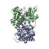
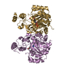
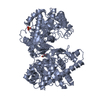
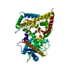
 Thermoplasma acidophilum (acidophilic)
 X-RAY DIFFRACTION /
  Authors
 Citation
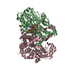
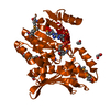
 Structure visualization
 Downloads & links
 Other downloads


 PDBj
PDBj

 Assembly
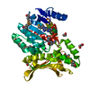


 Mass: 22.990 Da / Num. of mol.: 1 / Source method: obtained synthetically / Formula: Na
 Mass: 745.421 Da / Num. of mol.: 1 / Source method: obtained synthetically / Formula: C21H30N7O17P3
 Mass: 62.068 Da / Num. of mol.: 6 / Source method: obtained synthetically / Formula: C2H6O2
 Mass: 18.015 Da / Num. of mol.: 214 / Source method: isolated from a natural source / Formula: H2O
 Sample preparation
 / Beamline: BL9-2 / Wavelength: 0.91162,0.97927,0.97908
 Processing