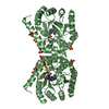
 Movie
Movie Controller

[English] 日本語
 Yorodumi
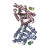
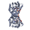
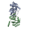
- PDB-3icw: Structure of a Circular Permutation on Lipase B from Candida Anta... -
+ Open data

- Basic information
| Entry | Database: PDB / ID: 3icw | ||||||
|---|---|---|---|---|---|---|---|
| Title | Structure of a Circular Permutation on Lipase B from Candida Antartica with Bound Suicide Inhibitor | ||||||
 Components Components | Lipase B | ||||||
 Keywords Keywords | HYDROLASE / CIRCULAR PERMUTATION / SUICIDE INHIBITION / Cleavage on pair of basic residues / Disulfide bond / Glycoprotein / Lipid degradation / Zymogen | ||||||
| Function / homology |  Function and homology information Function and homology informationtriacylglycerol lipase / triacylglycerol lipase activity / lipid catabolic process Similarity search - Function | ||||||
| Biological species |  Candida antarctica (fungus) Candida antarctica (fungus) | ||||||
| Method |  X-RAY DIFFRACTION / SYNCHROTRON / MOLECULAR REPLACEMENT / Resolution: 1.69 Å X-RAY DIFFRACTION / SYNCHROTRON / MOLECULAR REPLACEMENT / Resolution: 1.69 Å | ||||||
 Authors Authors | Horton, J.R. / Qian, Z. / Jia, D. / Lutz, S.A. / Cheng, X. | ||||||
 Citation Citation | Journal: J.Mol.Biol. / Year: 2009 Title: Structural redesign of lipase B from Candida antarctica by circular permutation and incremental truncation. Authors: Qian, Z. / Horton, J.R. / Cheng, X. / Lutz, S. | ||||||
| History |
|
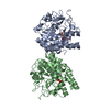
- Structure visualization
| Structure viewer | Molecule: MolmilJmol/JSmol |
|---|
- Downloads & links
-Download
| PDBx/mmCIF format | 3icw.cif.gz | 77.7 KB | Display | PDBx/mmCIF format |
|---|---|---|---|---|
| PDB format | pdb3icw.ent.gz | 57.3 KB | Display | PDB format |
| PDBx/mmJSON format | 3icw.json.gz | Tree view | PDBx/mmJSON format | |
| Others |  Other downloads Other downloads |
-Validation report
| Arichive directory | https://data.pdbj.org/pub/pdb/validation_reports/ic/3icwftp://data.pdbj.org/pub/pdb/validation_reports/ic/3icw | HTTPS FTP |
|---|
-Related structure data
| Related structure data | 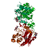 3icvC  1lbtS C: citing same article ( S: Starting model for refinement |
|---|---|
| Similar structure data |


-Links
 PDBj
PDBj
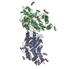
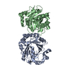
- Assembly
| Deposited unit | 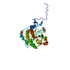
| ||||||||||||
|---|---|---|---|---|---|---|---|---|---|---|---|---|---|
| 1 | 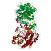
| ||||||||||||
| Unit cell |
| ||||||||||||
| Components on special symmetry positions |
|
-Components
| #1: Protein | Mass: 32899.070 Da / Num. of mol.: 1 Source method: isolated from a genetically manipulated source Source: (gene. exp.) Candida antarctica (fungus) / Production host: PICHIA PASTORIS (fungus) / References: UniProt: P41365, triacylglycerol lipase | ||||||||||
|---|---|---|---|---|---|---|---|---|---|---|---|
| #2: Sugar |   Type: D-saccharide, beta linking / Mass: 221.208 Da / Num. of mol.: 2 Type: D-saccharide, beta linking / Mass: 221.208 Da / Num. of mol.: 2Source method: isolated from a genetically manipulated source Formula: C8H15NO6 / References: triacylglycerol lipase #3: Chemical | ChemComp-PO4 / |   Mass: 94.971 Da / Num. of mol.: 1 / Source method: obtained synthetically / Formula: PO4 Mass: 94.971 Da / Num. of mol.: 1 / Source method: obtained synthetically / Formula: PO4#4: Chemical | ChemComp-MHH / |   Mass: 180.182 Da / Num. of mol.: 1 / Source method: obtained synthetically / Formula: C7H17O3P Mass: 180.182 Da / Num. of mol.: 1 / Source method: obtained synthetically / Formula: C7H17O3P#5: Water | ChemComp-HOH / |  Mass: 18.015 Da / Num. of mol.: 338 / Source method: isolated from a natural source / Formula: H2O Mass: 18.015 Da / Num. of mol.: 338 / Source method: isolated from a natural source / Formula: H2OHas protein modification | Y | Sequence details | THE RECOMBINANT PROTEIN IS A "CIRCULAR PERMUTATION" OF LIPASE B IN WHICH THE C-TERMINAL SEQUENCE ...THE RECOMBINAN | |
-Experimental details
-Experiment
| Experiment | Method: X-RAY DIFFRACTION / Number of used crystals: 1 |
|---|
- Sample preparation
| Crystal | Density Matthews: 3 Å3/Da / Density % sol: 59.03 % |
|---|---|
| Crystal grow | Temperature: 289 K / Method: vapor diffusion, hanging drop / pH: 4.5 Details: 20% PEG 3350, 0.2M SODIUM DIHYDROGEN PHOSPHATE, pH 4.5, VAPOR DIFFUSION, HANGING DROP, temperature 289K |
-Data collection
| Diffraction | Mean temperature: 173 K |
|---|---|
| Diffraction source | Source: SYNCHROTRON / Site: APS  / Beamline: 22-ID / Wavelength: 1 / Beamline: 22-ID / Wavelength: 1 |
| Detector | Type: MARMOSAIC 300 mm CCD / Detector: CCD / Date: Oct 20, 2006 |
| Radiation | Monochromator: SI(111) / Protocol: SINGLE WAVELENGTH / Monochromatic (M) / Laue (L): M / Scattering type: x-ray |
| Radiation wavelength | Wavelength: 1 Å / Relative weight: 1 |
| Reflection | Resolution: 1.69→27.8 Å / Num. obs: 44608 / % possible obs: 99.9 % / Observed criterion σ(I): -3 / Redundancy: 12.8 % / Biso Wilson estimate: 25.3 Å2 / Rmerge(I) obs: 0.121 / Net I/σ(I): 14.4 |
| Reflection shell | Resolution: 1.69→1.75 Å / Redundancy: 11.5 % / Rmerge(I) obs: 0.473 / Mean I/σ(I) obs: 4.2 / % possible all: 99.8 |
- Processing
| Software |
| ||||||||||||||||||||||||||||||||||||||||||||||||||||||||||||
|---|---|---|---|---|---|---|---|---|---|---|---|---|---|---|---|---|---|---|---|---|---|---|---|---|---|---|---|---|---|---|---|---|---|---|---|---|---|---|---|---|---|---|---|---|---|---|---|---|---|---|---|---|---|---|---|---|---|---|---|---|---|
| Refinement | Method to determine structure: MOLECULAR REPLACEMENT Starting model: PDB CODE 1LBT Resolution: 1.69→27.8 Å / Isotropic thermal model: ISOTROPIC / Cross valid method: THROUGHOUT / σ(F): 0 / Stereochemistry target values: Engh & Huber
| ||||||||||||||||||||||||||||||||||||||||||||||||||||||||||||
| Displacement parameters | Biso mean: 31.7 Å2
| ||||||||||||||||||||||||||||||||||||||||||||||||||||||||||||
| Refine analyze |
| ||||||||||||||||||||||||||||||||||||||||||||||||||||||||||||
| Refinement step | Cycle: LAST / Resolution: 1.69→27.8 Å
| ||||||||||||||||||||||||||||||||||||||||||||||||||||||||||||
| Refine LS restraints |
| ||||||||||||||||||||||||||||||||||||||||||||||||||||||||||||
| LS refinement shell | Resolution: 1.69→1.75 Å / Rfactor Rfree error: 0.018
|
