 Movie
Movie Controller

[English] 日本語
 Yorodumi
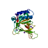
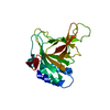
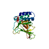
- PDB-3ici: Crystal structure of cyclophilin B in complex with calmegin fragment -
+ Open data

- Basic information
| Entry | Database: PDB / ID: 3ici | ||||||
|---|---|---|---|---|---|---|---|
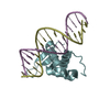
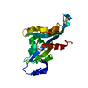
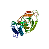
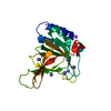
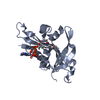
| Title | Crystal structure of cyclophilin B in complex with calmegin fragment | ||||||
 Components Components |
| ||||||
 Keywords Keywords | ISOMERASE / protein-protein complex / Endoplasmic reticulum / Glycoprotein / Rotamase / Chaperone / Lectin / Membrane / Phosphoprotein / Transmembrane | ||||||
| Function / homology |  Function and homology information Function and homology informationendoplasmic reticulum chaperone complex / host-mediated activation of viral genome replication / Collagen biosynthesis and modifying enzymes / host-mediated activation of viral process / positive regulation of multicellular organism growth / RNA polymerase binding / cyclosporin A binding / smooth endoplasmic reticulum / neutrophil chemotaxis / ERAD pathway ...endoplasmic reticulum chaperone complex / host-mediated activation of viral genome replication / Collagen biosynthesis and modifying enzymes / host-mediated activation of viral process / positive regulation of multicellular organism growth / RNA polymerase binding / cyclosporin A binding / smooth endoplasmic reticulum / neutrophil chemotaxis / ERAD pathway / peptidylprolyl isomerase / peptidyl-prolyl cis-trans isomerase activity / bone development / SARS-CoV-1 activates/modulates innate immune responses / : / melanosome / protein folding / protein stabilization / endoplasmic reticulum lumen / focal adhesion / calcium ion binding / endoplasmic reticulum membrane / perinuclear region of cytoplasm / endoplasmic reticulum / protein-containing complex / RNA binding / extracellular exosome / nucleoplasm / membrane / nucleus / cytosol / cytoplasm Similarity search - Function | ||||||
| Biological species |  Homo sapiens (human) Homo sapiens (human) | ||||||
| Method |  X-RAY DIFFRACTION / SYNCHROTRON / MOLECULAR REPLACEMENT / Resolution: 1.7 Å X-RAY DIFFRACTION / SYNCHROTRON / MOLECULAR REPLACEMENT / Resolution: 1.7 Å | ||||||
 Authors Authors | Kozlov, G. / Gehring, K. | ||||||
 Citation Citation | Journal: J.Biol.Chem. / Year: 2010 Title: Structural Basis of Cyclophilin B Binding by the Calnexin/Calreticulin P-domain. Authors: Kozlov, G. / Bastos-Aristizabal, S. / Maattanen, P. / Rosenauer, A. / Zheng, F. / Killikelly, A. / Trempe, J.F. / Thomas, D.Y. / Gehring, K. | ||||||
| History |
|
- Structure visualization
| Structure viewer | Molecule: MolmilJmol/JSmol |
|---|
- Downloads & links
-Download
| PDBx/mmCIF format | 3ici.cif.gz | 101.4 KB | Display | PDBx/mmCIF format |
|---|---|---|---|---|
| PDB format | pdb3ici.ent.gz | 75.4 KB | Display | PDB format |
| PDBx/mmJSON format | 3ici.json.gz | Tree view | PDBx/mmJSON format | |
| Others |  Other downloads Other downloads |
-Validation report
| Arichive directory | https://data.pdbj.org/pub/pdb/validation_reports/ic/3iciftp://data.pdbj.org/pub/pdb/validation_reports/ic/3ici | HTTPS FTP |
|---|
-Related structure data
| Related structure data |  3ichC 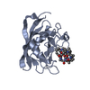 1cynS C: citing same article ( S: Starting model for refinement |
|---|---|
| Similar structure data |

-Links
 PDBj
PDBj


- Assembly
| Deposited unit | 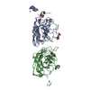
| ||||||||
|---|---|---|---|---|---|---|---|---|---|
| 1 | 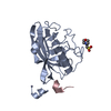
| ||||||||
| 2 | 
| ||||||||
| Unit cell |
|
-Components
| #1: Protein | Mass: 20735.787 Da / Num. of mol.: 2 / Fragment: UNP residues 34-216 Source method: isolated from a genetically manipulated source Source: (gene. exp.) Homo sapiens (human) / Gene: PPIB, CYPB / Plasmid: pGEX-6P-1 / Production host:  #2: Protein/peptide | | Mass: 4342.601 Da / Num. of mol.: 1 / Fragment: P-domain fragment: residues 317-350 Source method: isolated from a genetically manipulated source Source: (gene. exp.) PDB-D9N169, UniProt: E2RA18*PLUS#3: Chemical |   Mass: 65.409 Da / Num. of mol.: 2 / Source method: obtained synthetically / Formula: Zn Mass: 65.409 Da / Num. of mol.: 2 / Source method: obtained synthetically / Formula: Zn#4: Chemical | ChemComp-MES / |   Mass: 195.237 Da / Num. of mol.: 1 / Source method: obtained synthetically / Formula: C6H13NO4S / Comment: pH buffer*YM Mass: 195.237 Da / Num. of mol.: 1 / Source method: obtained synthetically / Formula: C6H13NO4S / Comment: pH buffer*YM#5: Water | ChemComp-HOH / |  Mass: 18.015 Da / Num. of mol.: 504 / Source method: isolated from a natural source / Formula: H2O Mass: 18.015 Da / Num. of mol.: 504 / Source method: isolated from a natural source / Formula: H2OSequence details | AUTHORS STATE THAT THE SEQUENCE OF ENTITY 2 (CHAIN C) CORRESPONDS TO RESIDUES 317-350 OF DOG ...AUTHORS STATE THAT THE SEQUENCE OF ENTITY 2 (CHAIN C) CORRESPOND | |
|---|
-Experimental details
-Experiment
| Experiment | Method: X-RAY DIFFRACTION / Number of used crystals: 1 |
|---|
- Sample preparation
| Crystal | Density Matthews: 2.12 Å3/Da / Density % sol: 41.9 % |
|---|---|
| Crystal grow | Temperature: 295 K / Method: vapor diffusion, hanging drop / pH: 7 Details: 22% w/v PEG 8000, 10mM ZnCl2, 0.1M Tris buffer, pH 7.0, VAPOR DIFFUSION, HANGING DROP, temperature 295K |
-Data collection
| Diffraction | Mean temperature: 100 K |
|---|---|
| Diffraction source | Source: SYNCHROTRON / Site: CHESS  / Beamline: A1 / Wavelength: 0.9995 Å / Beamline: A1 / Wavelength: 0.9995 Å |
| Detector | Type: ADSC QUANTUM 210 / Detector: CCD / Date: Nov 29, 2008 / Details: mirrors |
| Radiation | Monochromator: Si(111) channel / Protocol: SINGLE WAVELENGTH / Monochromatic (M) / Laue (L): M / Scattering type: x-ray |
| Radiation wavelength | Wavelength: 0.9995 Å / Relative weight: 1 |
| Reflection | Resolution: 1.7→50 Å / Num. obs: 38845 / % possible obs: 97.3 % / Observed criterion σ(I): 1 / Redundancy: 3.9 % / Rmerge(I) obs: 0.087 / Net I/σ(I): 18 |
| Reflection shell | Resolution: 1.7→1.76 Å / Redundancy: 3.4 % / Rmerge(I) obs: 0.263 / Mean I/σ(I) obs: 4.6 / Num. unique all: 2598 / % possible all: 95 |
- Processing
| Software |
| ||||||||||||||||||||||||||||||||||||||||||||||||||||||||||||||||||||||||||||||||||||||||||||||||||||
|---|---|---|---|---|---|---|---|---|---|---|---|---|---|---|---|---|---|---|---|---|---|---|---|---|---|---|---|---|---|---|---|---|---|---|---|---|---|---|---|---|---|---|---|---|---|---|---|---|---|---|---|---|---|---|---|---|---|---|---|---|---|---|---|---|---|---|---|---|---|---|---|---|---|---|---|---|---|---|---|---|---|---|---|---|---|---|---|---|---|---|---|---|---|---|---|---|---|---|---|---|---|
| Refinement | Method to determine structure: MOLECULAR REPLACEMENT Starting model: PDB entry 1CYN Resolution: 1.7→50 Å / Cor.coef. Fo:Fc: 0.947 / Cor.coef. Fo:Fc free: 0.909 / SU B: 2.316 / SU ML: 0.079 / Cross valid method: THROUGHOUT / σ(F): 0 / ESU R: 0.127 / ESU R Free: 0.128 / Stereochemistry target values: MAXIMUM LIKELIHOOD
| ||||||||||||||||||||||||||||||||||||||||||||||||||||||||||||||||||||||||||||||||||||||||||||||||||||
| Solvent computation | Ion probe radii: 0.8 Å / Shrinkage radii: 0.8 Å / VDW probe radii: 1.2 Å / Solvent model: MASK | ||||||||||||||||||||||||||||||||||||||||||||||||||||||||||||||||||||||||||||||||||||||||||||||||||||
| Displacement parameters | Biso mean: 16.291 Å2
| ||||||||||||||||||||||||||||||||||||||||||||||||||||||||||||||||||||||||||||||||||||||||||||||||||||
| Refinement step | Cycle: LAST / Resolution: 1.7→50 Å
| ||||||||||||||||||||||||||||||||||||||||||||||||||||||||||||||||||||||||||||||||||||||||||||||||||||
| Refine LS restraints |
| ||||||||||||||||||||||||||||||||||||||||||||||||||||||||||||||||||||||||||||||||||||||||||||||||||||
| LS refinement shell | Resolution: 1.7→1.75 Å / Total num. of bins used: 20
|
