 Movie
Movie Controller

[English] 日本語
 Yorodumi
- PDB-3iab: Crystal structure of RNase P /RNase MRP proteins Pop6, Pop7 in a ... -
+ Open data

- Basic information
| Entry | Database: PDB / ID: 3iab | ||||||
|---|---|---|---|---|---|---|---|
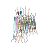
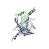
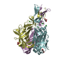
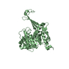
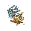
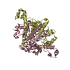
| Title | Crystal structure of RNase P /RNase MRP proteins Pop6, Pop7 in a complex with the P3 domain of RNase MRP RNA | ||||||
 Components Components |
| ||||||
 Keywords Keywords | HYDROLASE/RNA / RNase P / RNase MRP / Ribonuclease P / Ribonuclease MRP / Pop6 / Pop6p / Pop7 / Pop7p / P3 / NME1 / yeast / tRNA / pre-tRNA / rRNA / Ribozyme / PROTEIN-RNA COMPLEX / ALBA / heterodimer / Coiled coil / Hydrolase / Nucleus / rRNA processing / tRNA processing / Phosphoprotein / HYDROLASE-RNA COMPLEX | ||||||
| Function / homology |  Function and homology information Function and homology informationnuclear-transcribed mRNA catabolic process, RNase MRP-dependent / intronic box C/D snoRNA processing / nucleolar ribonuclease P complex / ribonuclease MRP complex / ribonuclease P / rRNA primary transcript binding / ribonuclease P activity / telomerase holoenzyme complex / tRNA 5'-leader removal / tRNA processing ...nuclear-transcribed mRNA catabolic process, RNase MRP-dependent / intronic box C/D snoRNA processing / nucleolar ribonuclease P complex / ribonuclease MRP complex / ribonuclease P / rRNA primary transcript binding / ribonuclease P activity / telomerase holoenzyme complex / tRNA 5'-leader removal / tRNA processing / maturation of 5.8S rRNA / rRNA processing / RNA binding / nucleus / cytosol Similarity search - Function | ||||||
| Biological species |  | ||||||
| Method |  X-RAY DIFFRACTION / SYNCHROTRON / SAD / Resolution: 2.7 Å X-RAY DIFFRACTION / SYNCHROTRON / SAD / Resolution: 2.7 Å | ||||||
 Authors Authors | Perederina, A. / Esakova, O.A. / Quan, C. / Khanova, E. / Krasilnikov, A.S. | ||||||
 Citation Citation | Journal: Embo J. / Year: 2010 Title: Eukaryotic ribonucleases P/MRP: the crystal structure of the P3 domain Authors: Perederina, A. / Esakova, O. / Quan, C. / Khanova, E. / Krasilnikov, A.S. | ||||||
| History |
|
- Structure visualization
| Structure viewer | Molecule: MolmilJmol/JSmol |
|---|
- Downloads & links
-Download
| PDBx/mmCIF format | 3iab.cif.gz | 93.1 KB | Display | PDBx/mmCIF format |
|---|---|---|---|---|
| PDB format | pdb3iab.ent.gz | 68.3 KB | Display | PDB format |
| PDBx/mmJSON format | 3iab.json.gz | Tree view | PDBx/mmJSON format | |
| Others |  Other downloads Other downloads |
-Validation report
| Arichive directory | https://data.pdbj.org/pub/pdb/validation_reports/ia/3iabftp://data.pdbj.org/pub/pdb/validation_reports/ia/3iab | HTTPS FTP |
|---|
-Related structure data
| Similar structure data |
|---|


-Links
 PDBj
PDBj


- Assembly
| Deposited unit | 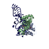
| ||||||||
|---|---|---|---|---|---|---|---|---|---|
| 1 |
| ||||||||
| 2 | 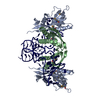
| ||||||||
| Unit cell |
| ||||||||
| Components on special symmetry positions |
|
-Components
| #1: Protein | Mass: 18440.576 Da / Num. of mol.: 1 / Fragment: Pop6 / Mutation: L141M Source method: isolated from a genetically manipulated source Details: Co-expression with Pop7 Source: (gene. exp.) Gene: POP6, YGR030C / Plasmid: 762 / Production host:  | ||||||
|---|---|---|---|---|---|---|---|
| #2: Protein | Mass: 15984.972 Da / Num. of mol.: 1 / Fragment: Pop7 / Mutation: none Source method: isolated from a genetically manipulated source Details: Co-expression with Pop6 Source: (gene. exp.) Gene: POP7, RPP2, YBR1219, YBR167C / Plasmid: 762 / Production host: | ||||||
| #3: RNA chain | Mass: 14828.875 Da / Num. of mol.: 1 / Fragment: P3 domain Mutation: circular permutation; A49C, A50C, U59G, U60G (yeast sequence numbering). See important note regarging nucleotide numbering in the model. Source method: obtained synthetically Details: In vitro transcription; circular permutation of naturally occurring sequence. References: ribonuclease P | ||||||
| #4: Chemical |   Mass: 65.409 Da / Num. of mol.: 3 / Source method: obtained synthetically / Formula: Zn Mass: 65.409 Da / Num. of mol.: 3 / Source method: obtained synthetically / Formula: Zn#5: Water | ChemComp-HOH / |  Mass: 18.015 Da / Num. of mol.: 50 / Source method: isolated from a natural source / Formula: H2O Mass: 18.015 Da / Num. of mol.: 50 / Source method: isolated from a natural source / Formula: H2OHas protein modification | Y | Sequence details | THE MODEL INCLUDES NUCLEOTIDES 29-50 AND 59-79 OF THE ORIGINAL YEAST P3 DOMAIN. THE CRYSTALLIZED ...THE MODEL INCLUDES NUCLEOTIDE | |
-Experimental details
-Experiment
| Experiment | Method: X-RAY DIFFRACTION / Number of used crystals: 1 |
|---|
- Sample preparation
| Crystal | Density Matthews: 3.12 Å3/Da / Density % sol: 60.55 % |
|---|---|
| Crystal grow | Temperature: 292 K / Method: vapor diffusion, sitting drop / pH: 7.8 Details: 2M Ammonium Sulfate, 200 mM Potassium Chloride, 2% PEG 400, 5% D-trehalose, 2 mM Zinc Chloride, 5 mM Magnesium Chloride, 100 mM HEPES-Na, pH 7.8, VAPOR DIFFUSION, SITTING DROP, temperature 292K |
-Data collection
| Diffraction | Mean temperature: 100 K |
|---|---|
| Diffraction source | Source: SYNCHROTRON / Site: NSLS  / Beamline: X29A / Wavelength: 0.9791 Å / Beamline: X29A / Wavelength: 0.9791 Å |
| Detector | Type: ADSC QUANTUM 315r / Detector: CCD / Date: Mar 28, 2009 / Details: mirrors |
| Radiation | Monochromator: Si(111) / Protocol: SINGLE WAVELENGTH / Monochromatic (M) / Laue (L): M / Scattering type: x-ray |
| Radiation wavelength | Wavelength: 0.9791 Å / Relative weight: 1 |
| Reflection | Resolution: 2.7→30 Å / Num. all: 17601 / Num. obs: 16350 / % possible obs: 92.9 % / Observed criterion σ(F): 0 / Observed criterion σ(I): 0 / Redundancy: 5 % / Biso Wilson estimate: 82.1 Å2 / Rmerge(I) obs: 0.057 |
| Reflection shell | Resolution: 2.7→2.76 Å / Redundancy: 2.7 % / Rmerge(I) obs: 0.374 / Mean I/σ(I) obs: 2 / Num. unique all: 934 / % possible all: 93.3 |
- Processing
| Software |
| |||||||||||||||||||||||||
|---|---|---|---|---|---|---|---|---|---|---|---|---|---|---|---|---|---|---|---|---|---|---|---|---|---|---|
| Refinement | Method to determine structure: SAD Starting model: none Resolution: 2.7→30 Å / Isotropic thermal model: Isotropic / Cross valid method: THROUGHOUT / σ(F): 0 / σ(I): 0 / Stereochemistry target values: Engh & Huber Details: Residues 1-3, 122-128 in Pop6 (chain A) and residues 1-13, 105-124 in Pop7 (chain B) are disordered
| |||||||||||||||||||||||||
| Displacement parameters | Biso mean: 89.9 Å2
| |||||||||||||||||||||||||
| Refine analyze | Luzzati coordinate error obs: 0.68 Å | |||||||||||||||||||||||||
| Refinement step | Cycle: LAST / Resolution: 2.7→30 Å
| |||||||||||||||||||||||||
| Refine LS restraints |
| |||||||||||||||||||||||||
| LS refinement shell | Resolution: 2.7→2.774 Å
|
