 Movie
Movie Controller

[English] 日本語
 Yorodumi
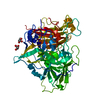
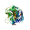
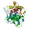
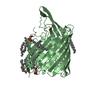
- PDB-1g6w: CRYSTAL STRUCTURE OF THE GLOBULAR REGION OF THE PRION PROTEIN URE... -
+ Open data

- Basic information
| Entry | Database: PDB / ID: 1g6w | ||||||
|---|---|---|---|---|---|---|---|
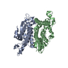
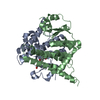
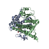
| Title | CRYSTAL STRUCTURE OF THE GLOBULAR REGION OF THE PRION PROTEIN URE2 FROM THE YEAST SACCAROMYCES CEREVISIAE | ||||||
 Components Components | URE2 PROTEIN | ||||||
 Keywords Keywords | STRUCTURAL GENOMICS / GST SUPERFAMILY | ||||||
| Function / homology |  Function and homology information Function and homology informationOxidoreductases; Acting on a sulfur group of donors; With a disulfide as acceptor / protein urmylation / glutathione peroxidase / regulation of nitrogen utilization / glutathione peroxidase activity / glutathione transferase activity / nitrate assimilation / phosphoprotein binding / transcription corepressor activity / intracellular protein localization ...Oxidoreductases; Acting on a sulfur group of donors; With a disulfide as acceptor / protein urmylation / glutathione peroxidase / regulation of nitrogen utilization / glutathione peroxidase activity / glutathione transferase activity / nitrate assimilation / phosphoprotein binding / transcription corepressor activity / intracellular protein localization / negative regulation of transcription by RNA polymerase II / cytoplasm Similarity search - Function | ||||||
| Biological species |  | ||||||
| Method |  X-RAY DIFFRACTION / SYNCHROTRON / MOLECULAR REPLACEMENT / Resolution: 2.5 Å X-RAY DIFFRACTION / SYNCHROTRON / MOLECULAR REPLACEMENT / Resolution: 2.5 Å | ||||||
 Authors Authors | Bousset, L. / Belrhali, H. / Janin, J. / Melki, R. / Morera, S. | ||||||
 Citation Citation | Journal: Structure / Year: 2001 Title: Structure of the globular region of the prion protein Ure2 from the yeast Saccharomyces cerevisiae. Authors: Bousset, L. / Belrhali, H. / Janin, J. / Melki, R. / Morera, S. #1: Journal: J.Biol.Chem. / Year: 1999Title: Structural Characterization of Saccharomyces cerevisiae Prion-like Protein Ure2 Authors: Thual, C. / Komar, A.A. / Bousset, L. / Fernandez-Bellot, E. / Cullin, C. / Melki, R. | ||||||
| History |
|
- Structure visualization
| Structure viewer | Molecule: MolmilJmol/JSmol |
|---|
- Downloads & links
-Download
| PDBx/mmCIF format | 1g6w.cif.gz | 207.6 KB | Display | PDBx/mmCIF format |
|---|---|---|---|---|
| PDB format | pdb1g6w.ent.gz | 168.2 KB | Display | PDB format |
| PDBx/mmJSON format | 1g6w.json.gz | Tree view | PDBx/mmJSON format | |
| Others |  Other downloads Other downloads |
-Validation report
| Arichive directory | https://data.pdbj.org/pub/pdb/validation_reports/g6/1g6wftp://data.pdbj.org/pub/pdb/validation_reports/g6/1g6w | HTTPS FTP |
|---|
-Related structure data


-Links
 PDBj
PDBj


- Assembly
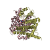
| Deposited unit | 
| ||||||||
|---|---|---|---|---|---|---|---|---|---|
| 1 | 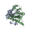
| ||||||||
| 2 | 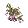
| ||||||||
| Unit cell |
|
-Components
| #1: Protein | Mass: 29966.104 Da / Num. of mol.: 4 / Fragment: GLOBULAR DOMAIN (RESIDUES 94-354) Source method: isolated from a genetically manipulated source Source: (gene. exp.) Gene: URE2 OR YNL229C OR N1165 / Plasmid: PET3 / Species (production host): Escherichia coli / Production host:  #2: Water | ChemComp-HOH / |  Mass: 18.015 Da / Num. of mol.: 286 / Source method: isolated from a natural source / Formula: H2O Mass: 18.015 Da / Num. of mol.: 286 / Source method: isolated from a natural source / Formula: H2O |
|---|
-Experimental details
-Experiment
| Experiment | Method: X-RAY DIFFRACTION / Number of used crystals: 1 |
|---|
- Sample preparation
| Crystal | Density Matthews: 2.28 Å3/Da / Density % sol: 46.04 % | ||||||||||||||||||||||||||||||
|---|---|---|---|---|---|---|---|---|---|---|---|---|---|---|---|---|---|---|---|---|---|---|---|---|---|---|---|---|---|---|---|
| Crystal grow | Temperature: 291 K / Method: vapor diffusion, hanging drop / pH: 8.5 Details: PEG 4000, CaCl2 , pH 8.5, VAPOR DIFFUSION, HANGING DROP, temperature 291K | ||||||||||||||||||||||||||||||
| Crystal | *PLUS Density % sol: 50 % | ||||||||||||||||||||||||||||||
| Crystal grow | *PLUS | ||||||||||||||||||||||||||||||
| Components of the solutions | *PLUS
|
-Data collection
| Diffraction | Mean temperature: 100 K |
|---|---|
| Diffraction source | Source: SYNCHROTRON / Site: ESRF  / Beamline: ID14-1 / Wavelength: 0.934 Å / Beamline: ID14-1 / Wavelength: 0.934 Å |
| Detector | Type: MARRESEARCH / Detector: CCD / Date: Dec 14, 1999 |
| Radiation | Monochromator: Asymmetric Laue C111 Diamond / Protocol: SINGLE WAVELENGTH / Monochromatic (M) / Laue (L): M / Scattering type: x-ray |
| Radiation wavelength | Wavelength: 0.934 Å / Relative weight: 1 |
| Reflection | Resolution: 2.5→20 Å / Num. all: 37704 / Num. obs: 36861 / % possible obs: 95 % / Observed criterion σ(F): 2 / Observed criterion σ(I): 1 / Redundancy: 3.8 % / Biso Wilson estimate: 44.454 Å2 / Rmerge(I) obs: 0.073 / Net I/σ(I): 5.7 |
| Reflection shell | Resolution: 2.5→2.64 Å / Redundancy: 3.5 % / Rmerge(I) obs: 0.291 / % possible all: 95 |
| Reflection | *PLUS Num. measured all: 277255 |
| Reflection shell | *PLUS % possible obs: 95 % / Mean I/σ(I) obs: 1.9 |
- Processing
| Software |
| ||||||||||||||||||||
|---|---|---|---|---|---|---|---|---|---|---|---|---|---|---|---|---|---|---|---|---|---|
| Refinement | Method to determine structure: MOLECULAR REPLACEMENT / Resolution: 2.5→20 Å / σ(F): 2 / σ(I): 1 / Stereochemistry target values: Engh & Huber
| ||||||||||||||||||||
| Refinement step | Cycle: LAST / Resolution: 2.5→20 Å
| ||||||||||||||||||||
| Refine LS restraints |
| ||||||||||||||||||||
| Software | *PLUS Name: CNS / Version: 1 / Classification: refinement | ||||||||||||||||||||
| Refinement | *PLUS Highest resolution: 2.5 Å / Lowest resolution: 20 Å / σ(F): 2 / % reflection Rfree: 5 % / Rfactor Rwork: 0.21 | ||||||||||||||||||||
| Solvent computation | *PLUS | ||||||||||||||||||||
| Displacement parameters | *PLUS Biso mean: 30.41 Å2 |
