 Movie
Movie Controller

[English] 日本語
 Yorodumi
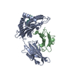
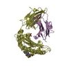
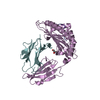
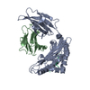
- PDB-3i6g: Newly identified epitope Mn2 from SARS-CoV M protein complexed wi... -
+ Open data

- Basic information
| Entry | Database: PDB / ID: 3i6g | ||||||
|---|---|---|---|---|---|---|---|
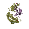
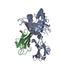
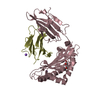
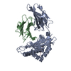
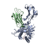
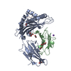
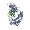
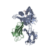
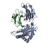
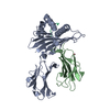
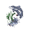
| Title | Newly identified epitope Mn2 from SARS-CoV M protein complexed withHLA-A*0201 | ||||||
 Components Components |
| ||||||
 Keywords Keywords | IMMUNE SYSTEM / HLA-A2 / SARS-CoV / membrane glycoprotein / Disulfide bond / Glycoprotein / Host-virus interaction / Immune response / Membrane / MHC I / Phosphoprotein / Transmembrane / Disease mutation / Glycation / Immunoglobulin domain / Pyrrolidone carboxylic acid / Secreted / Envelope protein / Golgi apparatus / Viral matrix protein / Virion | ||||||
| Function / homology |  Function and homology information Function and homology informationMaturation of protein M / Translation of Structural Proteins / Virion Assembly and Release / positive regulation of memory T cell activation / T cell mediated cytotoxicity directed against tumor cell target / TAP complex binding / Golgi medial cisterna / positive regulation of CD8-positive, alpha-beta T cell activation / CD8-positive, alpha-beta T cell activation / positive regulation of CD8-positive, alpha-beta T cell proliferation ...Maturation of protein M / Translation of Structural Proteins / Virion Assembly and Release / positive regulation of memory T cell activation / T cell mediated cytotoxicity directed against tumor cell target / TAP complex binding / Golgi medial cisterna / positive regulation of CD8-positive, alpha-beta T cell activation / CD8-positive, alpha-beta T cell activation / positive regulation of CD8-positive, alpha-beta T cell proliferation / CD8 receptor binding / antigen processing and presentation of exogenous peptide antigen via MHC class I / beta-2-microglobulin binding / endoplasmic reticulum exit site / antigen processing and presentation of endogenous peptide antigen via MHC class I via ER pathway, TAP-dependent / TAP binding / host cell Golgi membrane / protection from natural killer cell mediated cytotoxicity / antigen processing and presentation of endogenous peptide antigen via MHC class I via ER pathway, TAP-independent / antigen processing and presentation of endogenous peptide antigen via MHC class Ib / detection of bacterium / T cell receptor binding / SARS-CoV-1 targets host intracellular signalling and regulatory pathways / Attachment and Entry / negative regulation of receptor binding / early endosome lumen / Nef mediated downregulation of MHC class I complex cell surface expression / DAP12 interactions / transferrin transport / cellular response to iron ion / lumenal side of endoplasmic reticulum membrane / Endosomal/Vacuolar pathway / Antigen Presentation: Folding, assembly and peptide loading of class I MHC / peptide antigen assembly with MHC class II protein complex / antigen processing and presentation of exogenous protein antigen via MHC class Ib, TAP-dependent / cellular response to iron(III) ion / MHC class II protein complex / negative regulation of forebrain neuron differentiation / ER to Golgi transport vesicle membrane / peptide antigen assembly with MHC class I protein complex / regulation of erythrocyte differentiation / regulation of iron ion transport / response to molecule of bacterial origin / MHC class I peptide loading complex / HFE-transferrin receptor complex / T cell mediated cytotoxicity / antigen processing and presentation of endogenous peptide antigen via MHC class I / positive regulation of T cell cytokine production / antigen processing and presentation of exogenous peptide antigen via MHC class II / positive regulation of immune response / MHC class I protein complex / positive regulation of T cell activation / peptide antigen binding / positive regulation of receptor-mediated endocytosis / negative regulation of neurogenesis / positive regulation of T cell mediated cytotoxicity / cellular response to nicotine / multicellular organismal-level iron ion homeostasis / SARS-CoV-1 activates/modulates innate immune responses / Interferon alpha/beta signaling / Modulation by Mtb of host immune system / specific granule lumen / positive regulation of type II interferon production / phagocytic vesicle membrane / recycling endosome membrane / Immunoregulatory interactions between a Lymphoid and a non-Lymphoid cell / Interferon gamma signaling / negative regulation of epithelial cell proliferation / MHC class II protein complex binding / late endosome membrane / sensory perception of smell / antibacterial humoral response / positive regulation of cellular senescence / E3 ubiquitin ligases ubiquitinate target proteins / tertiary granule lumen / DAP12 signaling / T cell differentiation in thymus / ER-Phagosome pathway / T cell receptor signaling pathway / negative regulation of neuron projection development / protein refolding / early endosome membrane / protein homotetramerization / amyloid fibril formation / structural constituent of virion / intracellular iron ion homeostasis / learning or memory / defense response to Gram-positive bacterium / immune response / Amyloid fiber formation / endoplasmic reticulum lumen / Golgi membrane / signaling receptor binding / lysosomal membrane / innate immune response / external side of plasma membrane / focal adhesion / viral envelope / endoplasmic reticulum membrane / Neutrophil degranulation Similarity search - Function | ||||||
| Biological species |  Homo sapiens (human) Homo sapiens (human) | ||||||
| Method |  X-RAY DIFFRACTION / MOLECULAR REPLACEMENT / Resolution: 2.201 Å X-RAY DIFFRACTION / MOLECULAR REPLACEMENT / Resolution: 2.201 Å | ||||||
 Authors Authors | Liu, J. | ||||||
 Citation Citation | Journal: J.INFECT.DIS. / Year: 2010 Title: The membrane protein of severe acute respiratory syndrome coronavirus acts as a dominant immunogen revealed by a clustering region of novel functionally and structurally defined cytotoxic T-lymphocyte epitopes. Authors: Liu, J. / Sun, Y. / Qi, J. / Chu, F. / Wu, H. / Gao, F. / Li, T. / Yan, J. / Gao, G.F. | ||||||
| History |
|
- Structure visualization
| Structure viewer | Molecule: MolmilJmol/JSmol |
|---|
- Downloads & links
-Download
| PDBx/mmCIF format | 3i6g.cif.gz | 179.9 KB | Display | PDBx/mmCIF format |
|---|---|---|---|---|
| PDB format | pdb3i6g.ent.gz | 142.8 KB | Display | PDB format |
| PDBx/mmJSON format | 3i6g.json.gz | Tree view | PDBx/mmJSON format | |
| Others |  Other downloads Other downloads |
-Validation report
| Summary document | 3i6g_validation.pdf.gz | 453.5 KB | Display | wwPDB validaton report |
|---|---|---|---|---|
| Full document | 3i6g_full_validation.pdf.gz | 466.4 KB | Display | |
| Data in XML | 3i6g_validation.xml.gz | 35.7 KB | Display | |
| Data in CIF | 3i6g_validation.cif.gz | 51.5 KB | Display | |
| Arichive directory | https://data.pdbj.org/pub/pdb/validation_reports/i6/3i6gftp://data.pdbj.org/pub/pdb/validation_reports/i6/3i6g | HTTPS FTP |
-Related structure data
| Related structure data | 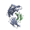 3i6kC  1zf1S S: Starting model for refinement C: citing same article ( |
|---|---|
| Similar structure data |


-Links
 PDBj
PDBj


- Assembly
| Deposited unit | 
| ||||||||
|---|---|---|---|---|---|---|---|---|---|
| 1 | 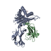
| ||||||||
| 2 | 
| ||||||||
| Unit cell |
|
-Components
| #1: Protein | Mass: 31854.203 Da / Num. of mol.: 2 / Fragment: UNP residues 25-299 Source method: isolated from a genetically manipulated source Source: (gene. exp.) Homo sapiens (human) / Gene: HLA-A, HLAA / Plasmid: pET21b / Production host:  #2: Protein | Mass: 11879.356 Da / Num. of mol.: 2 Source method: isolated from a genetically manipulated source Source: (gene. exp.) Homo sapiens (human) / Gene: B2M, CDABP0092, HDCMA22P / Plasmid: pGMT7 / Production host: #3: Protein/peptide | Mass: 1115.344 Da / Num. of mol.: 2 / Fragment: UNP residues 88-96 / Source method: obtained synthetically Details: Peptide Mn2 derived from membrane glycoprotein of SARS coronavirus TJF was synthesized. References: UniProt: P59596 #4: Water | ChemComp-HOH / |  Mass: 18.015 Da / Num. of mol.: 557 / Source method: isolated from a natural source / Formula: H2O Mass: 18.015 Da / Num. of mol.: 557 / Source method: isolated from a natural source / Formula: H2OHas protein modification | Y | |
|---|
-Experimental details
-Experiment
| Experiment | Method: X-RAY DIFFRACTION / Number of used crystals: 1 |
|---|
- Sample preparation
| Crystal | Density Matthews: 2.81 Å3/Da / Density % sol: 56.19 % |
|---|---|
| Crystal grow | Temperature: 277 K / Method: vapor diffusion, hanging drop / pH: 6 Details: 0.1M Ammonium acetate, 0.1M BIS-TRIS, pH6.0, 13-17% Polyethylene glycol 10,000, VAPOR DIFFUSION, HANGING DROP, temperature 277K |
-Data collection
| Diffraction | Mean temperature: 100 K |
|---|---|
| Diffraction source | Source: ROTATING ANODE / Type: RIGAKU MICROMAX-007 HF / Wavelength: 1.5478 Å |
| Detector | Type: RIGAKU RAXIS IV++ / Detector: IMAGE PLATE / Date: Sep 10, 2008 / Details: Mirror |
| Radiation | Protocol: SINGLE WAVELENGTH / Monochromatic (M) / Laue (L): M / Scattering type: x-ray |
| Radiation wavelength | Wavelength: 1.5478 Å / Relative weight: 1 |
| Reflection | Resolution: 2.2→27.3 Å / Num. obs: 50983 / % possible obs: 99.8 % / Observed criterion σ(F): 3 / Observed criterion σ(I): 3 / Redundancy: 3.5 % / Biso Wilson estimate: 39.3 Å2 / Rmerge(I) obs: 0.062 / Rsym value: 0.062 / Net I/σ(I): 31.682 |
| Reflection shell | Resolution: 2.2→2.28 Å / Redundancy: 3.5 % / Rmerge(I) obs: 0.319 / Mean I/σ(I) obs: 5.051 / Rsym value: 0.319 / % possible all: 100 |
- Processing
| Software |
| |||||||||||||||||||||||||||||||||||||||||||||||||||||||||||||||||||||||||||||||||||||||||||||||||||||||||||||||||||||||||||||||||||||
|---|---|---|---|---|---|---|---|---|---|---|---|---|---|---|---|---|---|---|---|---|---|---|---|---|---|---|---|---|---|---|---|---|---|---|---|---|---|---|---|---|---|---|---|---|---|---|---|---|---|---|---|---|---|---|---|---|---|---|---|---|---|---|---|---|---|---|---|---|---|---|---|---|---|---|---|---|---|---|---|---|---|---|---|---|---|---|---|---|---|---|---|---|---|---|---|---|---|---|---|---|---|---|---|---|---|---|---|---|---|---|---|---|---|---|---|---|---|---|---|---|---|---|---|---|---|---|---|---|---|---|---|---|---|---|
| Refinement | Method to determine structure: MOLECULAR REPLACEMENT Starting model: PDB ENTRY 1ZF1 Resolution: 2.201→27.254 Å / SU ML: 0.33 / σ(F): 1.34 / Stereochemistry target values: ML
| |||||||||||||||||||||||||||||||||||||||||||||||||||||||||||||||||||||||||||||||||||||||||||||||||||||||||||||||||||||||||||||||||||||
| Solvent computation | Shrinkage radii: 0.9 Å / VDW probe radii: 1.11 Å / Solvent model: FLAT BULK SOLVENT MODEL / Bsol: 50.145 Å2 / ksol: 0.356 e/Å3 | |||||||||||||||||||||||||||||||||||||||||||||||||||||||||||||||||||||||||||||||||||||||||||||||||||||||||||||||||||||||||||||||||||||
| Refinement step | Cycle: LAST / Resolution: 2.201→27.254 Å
| |||||||||||||||||||||||||||||||||||||||||||||||||||||||||||||||||||||||||||||||||||||||||||||||||||||||||||||||||||||||||||||||||||||
| Refine LS restraints |
| |||||||||||||||||||||||||||||||||||||||||||||||||||||||||||||||||||||||||||||||||||||||||||||||||||||||||||||||||||||||||||||||||||||
| LS refinement shell |
|
