 Movie
Movie Controller

[English] 日本語
 Yorodumi
- PDB-3i4z: Crystal structure of the dimethylallyl tryptophan synthase FgaPT2... -
+ Open data

- Basic information
| Entry | Database: PDB / ID: 3i4z | ||||||
|---|---|---|---|---|---|---|---|
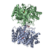
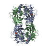
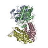
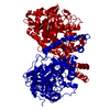
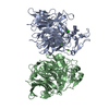
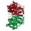
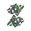
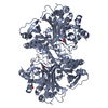
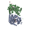
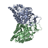
| Title | Crystal structure of the dimethylallyl tryptophan synthase FgaPT2 from Aspergillus fumigatus | ||||||
 Components Components | Tryptophan dimethylallyltransferase | ||||||
 Keywords Keywords | TRANSFERASE / prenyl transferase / PT barrel / Alkaloid metabolism | ||||||
| Function / homology |  Function and homology information Function and homology information4-dimethylallyltryptophan synthase / tryptophan dimethylallyltransferase activity / fumigaclavine C biosynthetic process / alkaloid biosynthetic process / prenyltransferase activity Similarity search - Function | ||||||
| Biological species |  | ||||||
| Method |  X-RAY DIFFRACTION / SYNCHROTRON / SAD / Resolution: 1.76 Å X-RAY DIFFRACTION / SYNCHROTRON / SAD / Resolution: 1.76 Å | ||||||
 Authors Authors | Schall, C. / Zocher, G. / Stehle, T. | ||||||
 Citation Citation | Journal: Proc.Natl.Acad.Sci.USA / Year: 2009 Title: The structure of dimethylallyl tryptophan synthase reveals a common architecture of aromatic prenyltransferases in fungi and bacteria Authors: Metzger, U. / Schall, C. / Zocher, G. / Unsoeld, I. / Stec, E. / Li, S.-M. / Heide, L. / Stehle, T. | ||||||
| History |
|
- Structure visualization
| Structure viewer | Molecule: MolmilJmol/JSmol |
|---|
- Downloads & links
-Download
| PDBx/mmCIF format | 3i4z.cif.gz | 375.9 KB | Display | PDBx/mmCIF format |
|---|---|---|---|---|
| PDB format | pdb3i4z.ent.gz | 307.4 KB | Display | PDB format |
| PDBx/mmJSON format | 3i4z.json.gz | Tree view | PDBx/mmJSON format | |
| Others |  Other downloads Other downloads |
-Validation report
| Arichive directory | https://data.pdbj.org/pub/pdb/validation_reports/i4/3i4zftp://data.pdbj.org/pub/pdb/validation_reports/i4/3i4z | HTTPS FTP |
|---|
-Related structure data

-Links
 PDBj
PDBj- Assembly
| Deposited unit | 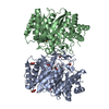
| ||||||||
|---|---|---|---|---|---|---|---|---|---|
| 1 |
| ||||||||
| Unit cell |
|
-Components
| #1: Protein | Mass: 52992.090 Da / Num. of mol.: 2 Source method: isolated from a genetically manipulated source Source: (gene. exp.)  References: UniProt: Q50EL0, 4-dimethylallyltryptophan synthase #2: Chemical | ChemComp-BU2 /   Mass: 90.121 Da / Num. of mol.: 8 / Source method: obtained synthetically / Formula: C4H10O2 Mass: 90.121 Da / Num. of mol.: 8 / Source method: obtained synthetically / Formula: C4H10O2#3: Chemical | ChemComp-GOL /   Mass: 92.094 Da / Num. of mol.: 4 / Source method: obtained synthetically / Formula: C3H8O3 Mass: 92.094 Da / Num. of mol.: 4 / Source method: obtained synthetically / Formula: C3H8O3#4: Water | ChemComp-HOH / |  Mass: 18.015 Da / Num. of mol.: 682 / Source method: isolated from a natural source / Formula: H2O Mass: 18.015 Da / Num. of mol.: 682 / Source method: isolated from a natural source / Formula: H2OSequence details | 1. RESIDUE SER443ALA IS A NATURAL VARIENT IN STRAIN: B5233 / ATCC 13073 OF DMAW_ASPFU ...1. RESIDUE SER443ALA IS A NATURAL VARIENT IN STRAIN: B5233 / ATCC 13073 OF DMAW_ASPFU (UNIPROTKB/SWISS-PROT Q50EL0). 2. AUTHOR CANNOT CLARIFY RESIDUES A 446(PRO) TO 458(LEU) BELONG TO EITHER CHAIN A OR CHAIN B. RESIDUES A 446(PRO) TO 458(LEU) WERE ASSIGNED TO CHAIN A TEMPORARY. | |
|---|
-Experimental details
-Experiment
| Experiment | Method: X-RAY DIFFRACTION / Number of used crystals: 2 |
|---|
- Sample preparation
| Crystal | Density Matthews: 2.34 Å3/Da / Density % sol: 47.52 % |
|---|---|
| Crystal grow | Temperature: 277 K / Method: vapor diffusion, hanging drop / pH: 7 Details: 26% 1,3-butanediol, 50mM sodium L-lactate, 100mM sodium MOPSO pH 7.0, 2mM DTT, vapour diffusion, hanging drop, temperature 277K, VAPOR DIFFUSION, HANGING DROP |
-Data collection
| Diffraction |
| ||||||||||||||||||||||||||||||||||||||||||||||||||||||||||||||||||||||||||||||||||||||||||||||||||||||||||||||||||||||||||||||
|---|---|---|---|---|---|---|---|---|---|---|---|---|---|---|---|---|---|---|---|---|---|---|---|---|---|---|---|---|---|---|---|---|---|---|---|---|---|---|---|---|---|---|---|---|---|---|---|---|---|---|---|---|---|---|---|---|---|---|---|---|---|---|---|---|---|---|---|---|---|---|---|---|---|---|---|---|---|---|---|---|---|---|---|---|---|---|---|---|---|---|---|---|---|---|---|---|---|---|---|---|---|---|---|---|---|---|---|---|---|---|---|---|---|---|---|---|---|---|---|---|---|---|---|---|---|---|---|
| Diffraction source |
| ||||||||||||||||||||||||||||||||||||||||||||||||||||||||||||||||||||||||||||||||||||||||||||||||||||||||||||||||||||||||||||||
| Detector |
| ||||||||||||||||||||||||||||||||||||||||||||||||||||||||||||||||||||||||||||||||||||||||||||||||||||||||||||||||||||||||||||||
| Radiation |
| ||||||||||||||||||||||||||||||||||||||||||||||||||||||||||||||||||||||||||||||||||||||||||||||||||||||||||||||||||||||||||||||
| Radiation wavelength |
| ||||||||||||||||||||||||||||||||||||||||||||||||||||||||||||||||||||||||||||||||||||||||||||||||||||||||||||||||||||||||||||||
| Reflection | Number: 536343 / Rmerge(I) obs: 0.134 / D res high: 2.7 Å / Num. obs: 52137 / % possible obs: 99.5 | ||||||||||||||||||||||||||||||||||||||||||||||||||||||||||||||||||||||||||||||||||||||||||||||||||||||||||||||||||||||||||||||
| Diffraction reflection shell |
| ||||||||||||||||||||||||||||||||||||||||||||||||||||||||||||||||||||||||||||||||||||||||||||||||||||||||||||||||||||||||||||||
| Reflection | Resolution: 1.76→24.71 Å / Num. obs: 98932 / % possible obs: 98.7 % / Observed criterion σ(F): 3 / Observed criterion σ(I): 0 / Redundancy: 3 % / Biso Wilson estimate: 29.8 Å2 / Rmerge(I) obs: 0.098 / Net I/σ(I): 14.14 | ||||||||||||||||||||||||||||||||||||||||||||||||||||||||||||||||||||||||||||||||||||||||||||||||||||||||||||||||||||||||||||||
| Reflection shell | Resolution: 1.76→1.81 Å / Rmerge(I) obs: 0.47 / Mean I/σ(I) obs: 2.2 / % possible all: 91.3 |

-Phasing
| Phasing | Method: SAD |
|---|
- Processing
| Software |
| |||||||||||||||||||||||||||||||||||||||||||||||||||||||||||||||||||||||||||
|---|---|---|---|---|---|---|---|---|---|---|---|---|---|---|---|---|---|---|---|---|---|---|---|---|---|---|---|---|---|---|---|---|---|---|---|---|---|---|---|---|---|---|---|---|---|---|---|---|---|---|---|---|---|---|---|---|---|---|---|---|---|---|---|---|---|---|---|---|---|---|---|---|---|---|---|---|
| Refinement | Method to determine structure: SAD / Resolution: 1.76→24.71 Å / Cor.coef. Fo:Fc: 0.971 / Cor.coef. Fo:Fc free: 0.959 / Occupancy max: 1 / Occupancy min: 0.3 / SU B: 4.333 / SU ML: 0.062 / Cross valid method: THROUGHOUT / σ(F): 0 / ESU R: 0.094 / ESU R Free: 0.093 / Stereochemistry target values: MAXIMUM LIKELIHOOD Details: HYDROGENS HAVE BEEN ADDED IN THE RIDING POSITIONS U VALUES: RESIDUAL ONLY
| |||||||||||||||||||||||||||||||||||||||||||||||||||||||||||||||||||||||||||
| Solvent computation | Ion probe radii: 0.8 Å / Shrinkage radii: 0.8 Å / VDW probe radii: 1.2 Å / Solvent model: MASK | |||||||||||||||||||||||||||||||||||||||||||||||||||||||||||||||||||||||||||
| Displacement parameters | Biso max: 76.49 Å2 / Biso mean: 26.02 Å2 / Biso min: 2 Å2
| |||||||||||||||||||||||||||||||||||||||||||||||||||||||||||||||||||||||||||
| Refinement step | Cycle: LAST / Resolution: 1.76→24.71 Å
| |||||||||||||||||||||||||||||||||||||||||||||||||||||||||||||||||||||||||||
| Refine LS restraints |
| |||||||||||||||||||||||||||||||||||||||||||||||||||||||||||||||||||||||||||
| LS refinement shell | Resolution: 1.76→1.805 Å / Total num. of bins used: 20
| |||||||||||||||||||||||||||||||||||||||||||||||||||||||||||||||||||||||||||
| Refinement TLS params. | Method: refined / Refine-ID: X-RAY DIFFRACTION
| |||||||||||||||||||||||||||||||||||||||||||||||||||||||||||||||||||||||||||
| Refinement TLS group |
|
