 Movie
Movie Controller

[English] 日本語
 Yorodumi
- PDB-3i3u: Crystal structure of lp_1913 protein from lactobacillus plantarum... -
+ Open data

- Basic information
| Entry | Database: PDB / ID: 3i3u | ||||||
|---|---|---|---|---|---|---|---|
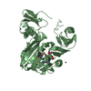
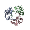
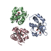
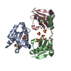
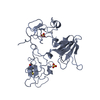
| Title | Crystal structure of lp_1913 protein from lactobacillus plantarum, northeast structural genomics Consortium target lpr140a | ||||||
 Components Components | uncharacterized protein lp_1913 | ||||||
 Keywords Keywords | structural genomics / unknown function / LACTOBACILLUS PLANTARUM / LP_1913 / PSI-2 / Protein Structure Initiative / Northeast Structural Genomics Consortium / NESG | ||||||
| Function / homology |  Function and homology information Function and homology information: / Rhodanese-like domain / Oxidized Rhodanese; domain 1 / Rhodanese Homology Domain / Rhodanese-like domain / Rhodanese domain profile. / Rhodanese-like domain superfamily / Rhodanese-like domain / 3-Layer(aba) Sandwich / Alpha Beta Similarity search - Domain/homology | ||||||
| Biological species |  Lactobacillus plantarum (bacteria) Lactobacillus plantarum (bacteria) | ||||||
| Method |  X-RAY DIFFRACTION / SYNCHROTRON / SAD / Resolution: 2.8 Å X-RAY DIFFRACTION / SYNCHROTRON / SAD / Resolution: 2.8 Å | ||||||
 Authors Authors | Seetharaman, J. / Chen, Y. / Forouhar, F. / Sahdev, S. / Janjua, H. / Xiao, R. / Ciccosanti, C. / Foote, E.L. / Acton, T.B. / Rost, B. ...Seetharaman, J. / Chen, Y. / Forouhar, F. / Sahdev, S. / Janjua, H. / Xiao, R. / Ciccosanti, C. / Foote, E.L. / Acton, T.B. / Rost, B. / Montelione, G.T. / Hunt, J.F. / Tong, L. / Northeast Structural Genomics Consortium (NESG) | ||||||
 Citation Citation | Journal: To be Published Title: Crystal structure of lp_1913 protein from lactobacillus plantarum, northeast structural genomics Consortium target lpr140a Authors: Seetharaman, J. / Chen, Y. / Forouhar, F. / Sahdev, S. / Janjua, H. / Xiao, R. / Ciccosanti, C. / Foote, E.L. / Acton, T.B. / Rost, B. / Montelione, G.T. / Hunt, J.F. / Tong, L. | ||||||
| History |
|
- Structure visualization
| Structure viewer | Molecule: MolmilJmol/JSmol |
|---|
- Downloads & links
-Download
| PDBx/mmCIF format | 3i3u.cif.gz | 109.4 KB | Display | PDBx/mmCIF format |
|---|---|---|---|---|
| PDB format | pdb3i3u.ent.gz | 85.6 KB | Display | PDB format |
| PDBx/mmJSON format | 3i3u.json.gz | Tree view | PDBx/mmJSON format | |
| Others |  Other downloads Other downloads |
-Validation report
| Arichive directory | https://data.pdbj.org/pub/pdb/validation_reports/i3/3i3uftp://data.pdbj.org/pub/pdb/validation_reports/i3/3i3u | HTTPS FTP |
|---|
-Related structure data
| Similar structure data | |
|---|---|
| Other databases |


-Links
 PDBj
PDBj- Assembly
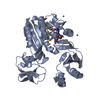
| Deposited unit | 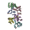
| ||||||||
|---|---|---|---|---|---|---|---|---|---|
| 1 | 
| ||||||||
| 2 | 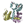
| ||||||||
| Unit cell |
|
-Components
| #1: Protein | Mass: 12324.717 Da / Num. of mol.: 6 Source method: isolated from a genetically manipulated source Source: (gene. exp.) Lactobacillus plantarum (bacteria) / Gene: lp_1913 / Plasmid: PET21 / Production host: #2: Water | ChemComp-HOH / |  Mass: 18.015 Da / Num. of mol.: 50 / Source method: isolated from a natural source / Formula: H2O Mass: 18.015 Da / Num. of mol.: 50 / Source method: isolated from a natural source / Formula: H2OHas protein modification | Y | |
|---|
-Experimental details
-Experiment
| Experiment | Method: X-RAY DIFFRACTION / Number of used crystals: 1 |
|---|
- Sample preparation
| Crystal | Density Matthews: 1.86 Å3/Da / Density % sol: 33.89 % |
|---|---|
| Crystal grow | Temperature: 293 K / Method: vapor diffusion, hanging drop / pH: 6 Details: NaBr 0.1M, MES 0.1M, PEG 8000 20%, pH 6.0, VAPOR DIFFUSION, HANGING DROP, temperature 293K |
-Data collection
| Diffraction | Mean temperature: 100 K |
|---|---|
| Diffraction source | Source: SYNCHROTRON / Site: NSLS  / Beamline: X4C / Wavelength: 0.979 Å / Beamline: X4C / Wavelength: 0.979 Å |
| Detector | Type: MAR CCD 165 mm / Detector: CCD / Date: Jun 2, 2009 |
| Radiation | Protocol: SINGLE WAVELENGTH / Monochromatic (M) / Laue (L): M / Scattering type: x-ray |
| Radiation wavelength | Wavelength: 0.979 Å / Relative weight: 1 |
| Reflection | Resolution: 2.5→50 Å / Num. all: 36176 / Num. obs: 36176 / % possible obs: 97.5 % / Observed criterion σ(F): 0 / Observed criterion σ(I): 0 / Redundancy: 1.9 % / Biso Wilson estimate: 0.1 Å2 / Rmerge(I) obs: 0.042 / Rsym value: 0.035 / Net I/σ(I): 13.9 |
| Reflection shell | Resolution: 2.5→2.59 Å / Redundancy: 1.9 % / Rmerge(I) obs: 0.213 / Num. unique all: 3389 / Rsym value: 0.198 / % possible all: 91 |
- Processing
| Software |
| ||||||||||||||||||||
|---|---|---|---|---|---|---|---|---|---|---|---|---|---|---|---|---|---|---|---|---|---|
| Refinement | Method to determine structure: SAD / Resolution: 2.8→26.12 Å / Rfactor Rfree error: 0.009 / Data cutoff high absF: 92884.23 / Data cutoff low absF: 0 / Isotropic thermal model: RESTRAINED / Cross valid method: THROUGHOUT / σ(F): 2 / Stereochemistry target values: Engh & Huber / Details: BULK SOLVENT MODEL USED
| ||||||||||||||||||||
| Solvent computation | Solvent model: FLAT MODEL / Bsol: 63.9606 Å2 / ksol: 0.4 e/Å3 | ||||||||||||||||||||
| Displacement parameters | Biso mean: 49.4 Å2
| ||||||||||||||||||||
| Refine analyze |
| ||||||||||||||||||||
| Refinement step | Cycle: LAST / Resolution: 2.8→26.12 Å
| ||||||||||||||||||||
| Refine LS restraints |
| ||||||||||||||||||||
| Refine LS restraints NCS | NCS model details: NONE | ||||||||||||||||||||
| LS refinement shell | Resolution: 2.8→2.98 Å / Rfactor Rfree error: 0.028 / Total num. of bins used: 6
| ||||||||||||||||||||
| Xplor file |
|
