 Movie
Movie Controller


+ Open data

- Basic information
| Entry | Database: PDB / ID: 3hxa | ||||||
|---|---|---|---|---|---|---|---|
| Title | Crystal Structure of DCoH1Thr51Ser | ||||||
 Components Components | Pterin-4-alpha-carbinolamine dehydratase | ||||||
 Keywords Keywords | LYASE / alpha and beta structure / Nucleus / Tetrahydrobiopterin biosynthesis | ||||||
| Function / homology |  Function and homology information Function and homology informationregulation of protein binding / Phenylalanine metabolism / 4a-hydroxytetrahydrobiopterin dehydratase / 4-alpha-hydroxytetrahydrobiopterin dehydratase activity / : / phenylalanine 4-monooxygenase activity / tetrahydrobiopterin biosynthetic process / transcription coactivator activity / positive regulation of DNA-templated transcription / nucleoplasm ...regulation of protein binding / Phenylalanine metabolism / 4a-hydroxytetrahydrobiopterin dehydratase / 4-alpha-hydroxytetrahydrobiopterin dehydratase activity / : / phenylalanine 4-monooxygenase activity / tetrahydrobiopterin biosynthetic process / transcription coactivator activity / positive regulation of DNA-templated transcription / nucleoplasm / identical protein binding / nucleus / cytosol Similarity search - Function | ||||||
| Biological species |  | ||||||
| Method |  X-RAY DIFFRACTION / SYNCHROTRON / MOLECULAR REPLACEMENT / molecular replacement / Resolution: 1.8 Å X-RAY DIFFRACTION / SYNCHROTRON / MOLECULAR REPLACEMENT / molecular replacement / Resolution: 1.8 Å | ||||||
 Authors Authors | Rho, H.Y. / Jones, C.N. / Rose, R.B. | ||||||
 Citation Citation | Journal: To be Published Title: Crystal Structure of DCoH1Thr51Ser Authors: Rho, H.Y. / Jones, C.N. / Rose, R.B. #1: Journal: Science / Year: 1995Title: Crystal structure of DCoH, a bifunctional, protein-binding transcriptional coactivator Authors: Endrizzi, J.A. / Cronk, J.D. / Wang, W. / Crabtree, G.R. / Alber, T. #2: Journal: Biochemistry / Year: 2004Title: Biochemical and structural basis for partially redundant enzymatic and transcriptional functions of DCoH and DCoH2 Authors: Rose, R.B. / Pullen, K.E. / Bayle, J.H. / Crabtree, G.R. / Alber, T. | ||||||
| History |
|
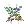
- Structure visualization
| Structure viewer | Molecule: MolmilJmol/JSmol |
|---|
- Downloads & links
-Download
| PDBx/mmCIF format | 3hxa.cif.gz | 179.8 KB | Display | PDBx/mmCIF format |
|---|---|---|---|---|
| PDB format | pdb3hxa.ent.gz | 145.4 KB | Display | PDB format |
| PDBx/mmJSON format | 3hxa.json.gz | Tree view | PDBx/mmJSON format | |
| Others |  Other downloads Other downloads |
-Validation report
| Arichive directory | https://data.pdbj.org/pub/pdb/validation_reports/hx/3hxaftp://data.pdbj.org/pub/pdb/validation_reports/hx/3hxa | HTTPS FTP |
|---|
-Related structure data
| Similar structure data |
|---|


-Links
 PDBj
PDBj

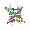
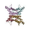
- Assembly
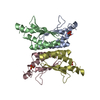
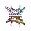
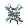
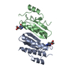
| Deposited unit | 
| ||||||||
|---|---|---|---|---|---|---|---|---|---|
| 1 | 
| ||||||||
| 2 | 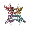
| ||||||||
| Unit cell |
|
-Components
| #1: Protein | Mass: 12003.576 Da / Num. of mol.: 8 / Mutation: T51S Source method: isolated from a genetically manipulated source Details: GST-FUSION / Source: (gene. exp.)  References: UniProt: P61459, 4a-hydroxytetrahydrobiopterin dehydratase #2: Chemical | ChemComp-SO4 /   Mass: 96.063 Da / Num. of mol.: 8 / Source method: obtained synthetically / Formula: SO4 Mass: 96.063 Da / Num. of mol.: 8 / Source method: obtained synthetically / Formula: SO4#3: Chemical | ChemComp-GOL /   Mass: 92.094 Da / Num. of mol.: 8 / Source method: obtained synthetically / Formula: C3H8O3 Mass: 92.094 Da / Num. of mol.: 8 / Source method: obtained synthetically / Formula: C3H8O3#4: Water | ChemComp-HOH / |  Mass: 18.015 Da / Num. of mol.: 524 / Source method: isolated from a natural source / Formula: H2O Mass: 18.015 Da / Num. of mol.: 524 / Source method: isolated from a natural source / Formula: H2O |
|---|
-Experimental details
-Experiment
| Experiment | Method: X-RAY DIFFRACTION / Number of used crystals: 1 |
|---|
- Sample preparation
| Crystal | Density Matthews: 3.14 Å3/Da / Density % sol: 60.78 % |
|---|---|
| Crystal grow | Temperature: 291 K / Method: hanging drop / pH: 7.5 Details: HEPES, Ammonium Sulfate, PEG 200, Glycerol, pH 7.5, Hanging drop, temperature 291K |
-Data collection
| Diffraction | Mean temperature: 100 K | ||||||||||||||||||||||||||||||||||||||||||||||||||||||||||||||||||||||||||||||||||||||||
|---|---|---|---|---|---|---|---|---|---|---|---|---|---|---|---|---|---|---|---|---|---|---|---|---|---|---|---|---|---|---|---|---|---|---|---|---|---|---|---|---|---|---|---|---|---|---|---|---|---|---|---|---|---|---|---|---|---|---|---|---|---|---|---|---|---|---|---|---|---|---|---|---|---|---|---|---|---|---|---|---|---|---|---|---|---|---|---|---|---|
| Diffraction source | Source: SYNCHROTRON / Site: APS  / Beamline: 22-ID / Beamline: 22-ID | ||||||||||||||||||||||||||||||||||||||||||||||||||||||||||||||||||||||||||||||||||||||||
| Detector | Type: MARMOSAIC 300 mm CCD / Detector: CCD / Date: Jul 1, 2006 | ||||||||||||||||||||||||||||||||||||||||||||||||||||||||||||||||||||||||||||||||||||||||
| Radiation | Protocol: SINGLE WAVELENGTH / Monochromatic (M) / Laue (L): M / Scattering type: x-ray | ||||||||||||||||||||||||||||||||||||||||||||||||||||||||||||||||||||||||||||||||||||||||
| Radiation wavelength | Relative weight: 1 | ||||||||||||||||||||||||||||||||||||||||||||||||||||||||||||||||||||||||||||||||||||||||
| Reflection | Resolution: 1.8→81.54 Å / Num. obs: 111375 / % possible obs: 99.1 % / Redundancy: 9.7 % / Rmerge(I) obs: 0.118 / Rsym value: 0.118 / Net I/σ(I): 2.958 | ||||||||||||||||||||||||||||||||||||||||||||||||||||||||||||||||||||||||||||||||||||||||
| Reflection shell |
|
-Phasing
| Phasing | Method: molecular replacement |
|---|
- Processing
| Software |
| ||||||||||||||||||||||||||||
|---|---|---|---|---|---|---|---|---|---|---|---|---|---|---|---|---|---|---|---|---|---|---|---|---|---|---|---|---|---|
| Refinement | Method to determine structure: MOLECULAR REPLACEMENT / Resolution: 1.8→50 Å / Occupancy max: 1 / Occupancy min: 0.5 / σ(F): 0
| ||||||||||||||||||||||||||||
| Solvent computation | Bsol: 57.222 Å2 | ||||||||||||||||||||||||||||
| Displacement parameters | Biso max: 73.94 Å2 / Biso mean: 32.07 Å2 / Biso min: 11.65 Å2
| ||||||||||||||||||||||||||||
| Refinement step | Cycle: LAST / Resolution: 1.8→50 Å
| ||||||||||||||||||||||||||||
| Refine LS restraints |
| ||||||||||||||||||||||||||||
| Xplor file |
|
