 Movie
Movie Controller

[English] 日本語
 Yorodumi
- PDB-1ru0: Crystal structure of DCoH2, a paralog of DCoH, the Dimerization C... -
+ Open data

- Basic information
| Entry | Database: PDB / ID: 1ru0 | ||||||
|---|---|---|---|---|---|---|---|
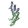
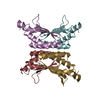
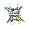
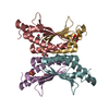
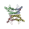
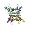
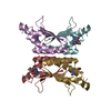
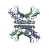
| Title | Crystal structure of DCoH2, a paralog of DCoH, the Dimerization Cofactor of HNF-1 | ||||||
 Components Components | DcoH-like protein DCoHm | ||||||
 Keywords Keywords | LYASE / alpha and beta structure | ||||||
| Function / homology |  Function and homology information Function and homology information4a-hydroxytetrahydrobiopterin dehydratase / 4-alpha-hydroxytetrahydrobiopterin dehydratase activity / phenylalanine 4-monooxygenase activity / tetrahydrobiopterin biosynthetic process / positive regulation of DNA-templated transcription / mitochondrion / identical protein binding / nucleus Similarity search - Function | ||||||
| Biological species |  | ||||||
| Method |  X-RAY DIFFRACTION / SYNCHROTRON / MOLECULAR REPLACEMENT / Resolution: 1.6 Å X-RAY DIFFRACTION / SYNCHROTRON / MOLECULAR REPLACEMENT / Resolution: 1.6 Å | ||||||
 Authors Authors | Rose, R.B. / Pullen, K.E. / Bayle, J.H. / Crabtree, G.R. / Alber, T. | ||||||
 Citation Citation | Journal: Biochemistry / Year: 2004 Title: Biochemical and structural basis for partially redundant enzymatic and transcriptional functions of DCoH and DCoH2 Authors: Rose, R.B. / Pullen, K.E. / Bayle, J.H. / Crabtree, G.R. / Alber, T. #1: Journal: Nat.Struct.Biol. / Year: 2000Title: Structural basis of dimerization, coactivator recognition and MODY3 mutations in HNF-1alpha Authors: Rose, R.B. / Bayle, J.H. / Endrizzi, J.A. / Cronk, J.D. / Crabtree, G.R. / Alber, T. | ||||||
| History |
|
- Structure visualization
| Structure viewer | Molecule: MolmilJmol/JSmol |
|---|
- Downloads & links
-Download
| PDBx/mmCIF format | 1ru0.cif.gz | 55 KB | Display | PDBx/mmCIF format |
|---|---|---|---|---|
| PDB format | pdb1ru0.ent.gz | 39.3 KB | Display | PDB format |
| PDBx/mmJSON format | 1ru0.json.gz | Tree view | PDBx/mmJSON format | |
| Others |  Other downloads Other downloads |
-Validation report
| Arichive directory | https://data.pdbj.org/pub/pdb/validation_reports/ru/1ru0ftp://data.pdbj.org/pub/pdb/validation_reports/ru/1ru0 | HTTPS FTP |
|---|
-Related structure data
| Related structure data |  1dcoS S: Starting model for refinement |
|---|---|
| Similar structure data |


-Links
 PDBj
PDBj
- Assembly
| Deposited unit | 
| |||||||||
|---|---|---|---|---|---|---|---|---|---|---|
| 1 | 
| |||||||||
| Unit cell |
| |||||||||
| Components on special symmetry positions |
| |||||||||
| Details | A dimer is in the asymmetric unit, which interacts with HNF-1. DCoH2 is a tetramer in solution. The tetramer is generated by rotating the dimer around the crystallographic two-fold axis: -x, -x+y, -z+1/3. |
-Components
| #1: Protein | Mass: 11899.393 Da / Num. of mol.: 2 Source method: isolated from a genetically manipulated source Source: (gene. exp.)  References: UniProt: Q9CZL5, 4a-hydroxytetrahydrobiopterin dehydratase #2: Water | ChemComp-HOH / |  Mass: 18.015 Da / Num. of mol.: 163 / Source method: isolated from a natural source / Formula: H2O Mass: 18.015 Da / Num. of mol.: 163 / Source method: isolated from a natural source / Formula: H2O |
|---|
-Experimental details
-Experiment
| Experiment | Method: X-RAY DIFFRACTION / Number of used crystals: 1 |
|---|
- Sample preparation
| Crystal | Density Matthews: 2.36 Å3/Da / Density % sol: 47.92 % |
|---|---|
| Crystal grow | Temperature: 277 K / Method: vapor diffusion, hanging drop / pH: 7.5 Details: isopropanol, PEG 4000, Na Hepes, pH 7.5, VAPOR DIFFUSION, HANGING DROP, temperature 277K |
-Data collection
| Diffraction | Mean temperature: 100 K |
|---|---|
| Diffraction source | Source: SYNCHROTRON / Site: ALS  / Beamline: 5.0.2 / Wavelength: 1 Å / Beamline: 5.0.2 / Wavelength: 1 Å |
| Detector | Type: ADSC QUANTUM 4 / Detector: CCD / Date: May 1, 2000 |
| Radiation | Monochromator: NULL / Protocol: SINGLE WAVELENGTH / Monochromatic (M) / Laue (L): M / Scattering type: x-ray |
| Radiation wavelength | Wavelength: 1 Å / Relative weight: 1 |
| Reflection | Resolution: 1.6→50 Å / Num. all: 30110 / Num. obs: 30110 / % possible obs: 99.9 % / Observed criterion σ(F): 2 / Observed criterion σ(I): 0 / Redundancy: 4.7 % / Biso Wilson estimate: 18.5 Å2 / Rsym value: 0.066 / Net I/σ(I): 12.5 |
| Reflection shell | Resolution: 1.6→1.69 Å / Redundancy: 3.3 % / Mean I/σ(I) obs: 3.8 / Num. unique all: 4325 / Rsym value: 0.253 / % possible all: 99.9 |
- Processing
| Software |
| |||||||||||||||||||||||||
|---|---|---|---|---|---|---|---|---|---|---|---|---|---|---|---|---|---|---|---|---|---|---|---|---|---|---|
| Refinement | Method to determine structure: MOLECULAR REPLACEMENT Starting model: pdb entry 1DCO Resolution: 1.6→50 Å / Isotropic thermal model: anisotropic / Cross valid method: THROUGHOUT / σ(F): 0 / Stereochemistry target values: Engh & Huber
| |||||||||||||||||||||||||
| Solvent computation | Bsol: 89.3426 Å2 / ksol: 0.414439 e/Å3 | |||||||||||||||||||||||||
| Displacement parameters | Biso mean: 21.4 Å2
| |||||||||||||||||||||||||
| Refine analyze |
| |||||||||||||||||||||||||
| Refinement step | Cycle: LAST / Resolution: 1.6→50 Å
| |||||||||||||||||||||||||
| Refine LS restraints |
| |||||||||||||||||||||||||
| LS refinement shell | Resolution: 1.6→1.66 Å
| |||||||||||||||||||||||||
| Xplor file |
|
