 Movie
Movie Controller

[English] 日本語
 Yorodumi
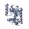
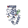
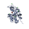
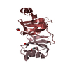
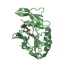
- PDB-3hw5: crystal structure of avian influenza virus PA_N in complex with AMP -
+ Open data

- Basic information
| Entry | Database: PDB / ID: 3hw5 | ||||||
|---|---|---|---|---|---|---|---|
| Title | crystal structure of avian influenza virus PA_N in complex with AMP | ||||||
 Components Components | Polymerase acidic protein | ||||||
 Keywords Keywords | HYDROLASE / avian influenza virus / PA_N / AMP / Phosphoprotein | ||||||
| Function / homology |  Function and homology information Function and homology informationcap snatching / symbiont-mediated suppression of host mRNA transcription via inhibition of RNA polymerase II activity / endonuclease activity / Hydrolases; Acting on ester bonds / host cell cytoplasm / symbiont-mediated suppression of host gene expression / viral translational frameshifting / viral RNA genome replication / hydrolase activity / DNA-templated transcription ...cap snatching / symbiont-mediated suppression of host mRNA transcription via inhibition of RNA polymerase II activity / endonuclease activity / Hydrolases; Acting on ester bonds / host cell cytoplasm / symbiont-mediated suppression of host gene expression / viral translational frameshifting / viral RNA genome replication / hydrolase activity / DNA-templated transcription / host cell nucleus / RNA binding / metal ion binding Similarity search - Function | ||||||
| Biological species |   Influenza A virus Influenza A virus | ||||||
| Method |  X-RAY DIFFRACTION / SYNCHROTRON / MOLECULAR REPLACEMENT / Resolution: 1.81 Å X-RAY DIFFRACTION / SYNCHROTRON / MOLECULAR REPLACEMENT / Resolution: 1.81 Å | ||||||
 Authors Authors | Zhao, C. / Lou, Z. / Guo, Y. / Ma, M. / Chen, Y. / Liang, S. / Rao, Z. | ||||||
 Citation Citation | Journal: J.Virol. / Year: 2009 Title: Nucleoside monophosphate complex structures of the endonuclease domain from the influenza virus polymerase PA subunit reveal the substrate binding site inside the catalytic center Authors: Zhao, C. / Lou, Z. / Guo, Y. / Ma, M. / Chen, Y. / Liang, S. / Zhang, L. / Chen, S. / Li, X. / Liu, Y. / Bartlam, M. / Rao, Z. | ||||||
| History |
|
- Structure visualization
| Structure viewer | Molecule: MolmilJmol/JSmol |
|---|
- Downloads & links
-Download
| PDBx/mmCIF format | 3hw5.cif.gz | 178.3 KB | Display | PDBx/mmCIF format |
|---|---|---|---|---|
| PDB format | pdb3hw5.ent.gz | 137.3 KB | Display | PDB format |
| PDBx/mmJSON format | 3hw5.json.gz | Tree view | PDBx/mmJSON format | |
| Others |  Other downloads Other downloads |
-Validation report
| Arichive directory | https://data.pdbj.org/pub/pdb/validation_reports/hw/3hw5ftp://data.pdbj.org/pub/pdb/validation_reports/hw/3hw5 | HTTPS FTP |
|---|
-Related structure data
| Related structure data |  3hw3C  3hw4C  3hw6C  3ebjS C: citing same article ( S: Starting model for refinement |
|---|---|
| Similar structure data |


-Links
 PDBj
PDBj


- Assembly
| Deposited unit | 
| ||||||||
|---|---|---|---|---|---|---|---|---|---|
| 1 | 
| ||||||||
| 2 | 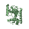
| ||||||||
| 3 | 
| ||||||||
| 4 | 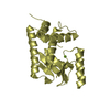
| ||||||||
| Unit cell |
|
-Components
| #1: Protein | Mass: 30324.307 Da / Num. of mol.: 4 / Fragment: residues in UNP 1- 256 / Mutation: V201I Source method: isolated from a genetically manipulated source Source: (gene. exp.) Influenza A virus (A/Goose/Guangdong/1/96(H5N1))Gene: PA / Plasmid: pGEX-6p-1 / Production host:  #2: Chemical | ChemComp-MG /   Mass: 24.305 Da / Num. of mol.: 4 / Source method: obtained synthetically / Formula: Mg Mass: 24.305 Da / Num. of mol.: 4 / Source method: obtained synthetically / Formula: Mg#3: Chemical | ChemComp-AMP / |   Mass: 347.221 Da / Num. of mol.: 1 / Source method: obtained synthetically / Formula: C10H14N5O7P / Comment: AMP*YM Mass: 347.221 Da / Num. of mol.: 1 / Source method: obtained synthetically / Formula: C10H14N5O7P / Comment: AMP*YM#4: Water | ChemComp-HOH / |  Mass: 18.015 Da / Num. of mol.: 632 / Source method: isolated from a natural source / Formula: H2O Mass: 18.015 Da / Num. of mol.: 632 / Source method: isolated from a natural source / Formula: H2OSequence details | THE AUTHORS BELIEVE THAT ILE 201 IS CORRECT AND IT IS NATURAL MUTANT. | |
|---|
-Experimental details
-Experiment
| Experiment | Method: X-RAY DIFFRACTION / Number of used crystals: 1 |
|---|
- Sample preparation
| Crystal | Density Matthews: 2.1 Å3/Da / Density % sol: 42.4 % |
|---|---|
| Crystal grow | Temperature: 291 K / Method: vapor diffusion, sitting drop / pH: 6.5 Details: 17.5% PEG3350(w/v), 100mM MgAc, 100mM MES, pH 6.5, VAPOR DIFFUSION, SITTING DROP, temperature 291K |
-Data collection
| Diffraction | Mean temperature: 100 K |
|---|---|
| Diffraction source | Source: SYNCHROTRON / Site: Photon Factory  / Beamline: BL-17A / Wavelength: 1 Å / Beamline: BL-17A / Wavelength: 1 Å |
| Detector | Type: ADSC QUANTUM 270 / Detector: CCD / Date: Mar 28, 2009 |
| Radiation | Protocol: SINGLE WAVELENGTH / Monochromatic (M) / Laue (L): M / Scattering type: x-ray |
| Radiation wavelength | Wavelength: 1 Å / Relative weight: 1 |
| Reflection | Resolution: 1.8→50 Å / Num. all: 63099 / Num. obs: 65112 / % possible obs: 96 % / Observed criterion σ(F): 0 / Rmerge(I) obs: 0.054 |
| Reflection shell | Resolution: 1.8→1.9 Å / Rmerge(I) obs: 0.128 / Num. unique all: 7817 / % possible all: 96 |
- Processing
| Software |
| ||||||||||||||||||||||||||||||||||||||||||||||||||||||||||||||||||||||||||||||||||||||||||||||||||||||||||||||||||||||||||||||||||||||||||||||||||||||||||||||||||||||||||
|---|---|---|---|---|---|---|---|---|---|---|---|---|---|---|---|---|---|---|---|---|---|---|---|---|---|---|---|---|---|---|---|---|---|---|---|---|---|---|---|---|---|---|---|---|---|---|---|---|---|---|---|---|---|---|---|---|---|---|---|---|---|---|---|---|---|---|---|---|---|---|---|---|---|---|---|---|---|---|---|---|---|---|---|---|---|---|---|---|---|---|---|---|---|---|---|---|---|---|---|---|---|---|---|---|---|---|---|---|---|---|---|---|---|---|---|---|---|---|---|---|---|---|---|---|---|---|---|---|---|---|---|---|---|---|---|---|---|---|---|---|---|---|---|---|---|---|---|---|---|---|---|---|---|---|---|---|---|---|---|---|---|---|---|---|---|---|---|---|---|---|---|
| Refinement | Method to determine structure: MOLECULAR REPLACEMENT Starting model: 3EBJ Resolution: 1.81→50 Å / Cor.coef. Fo:Fc: 0.948 / Cor.coef. Fo:Fc free: 0.922 / SU B: 3.261 / SU ML: 0.105 / Cross valid method: THROUGHOUT / ESU R: 0.171 / ESU R Free: 0.164 / Stereochemistry target values: MAXIMUM LIKELIHOOD / Details: HYDROGENS HAVE BEEN ADDED IN THE RIDING POSITIONS
| ||||||||||||||||||||||||||||||||||||||||||||||||||||||||||||||||||||||||||||||||||||||||||||||||||||||||||||||||||||||||||||||||||||||||||||||||||||||||||||||||||||||||||
| Solvent computation | Ion probe radii: 0.8 Å / Shrinkage radii: 0.8 Å / VDW probe radii: 1.4 Å / Solvent model: MASK | ||||||||||||||||||||||||||||||||||||||||||||||||||||||||||||||||||||||||||||||||||||||||||||||||||||||||||||||||||||||||||||||||||||||||||||||||||||||||||||||||||||||||||
| Displacement parameters | Biso mean: 29.176 Å2
| ||||||||||||||||||||||||||||||||||||||||||||||||||||||||||||||||||||||||||||||||||||||||||||||||||||||||||||||||||||||||||||||||||||||||||||||||||||||||||||||||||||||||||
| Refinement step | Cycle: LAST / Resolution: 1.81→50 Å
| ||||||||||||||||||||||||||||||||||||||||||||||||||||||||||||||||||||||||||||||||||||||||||||||||||||||||||||||||||||||||||||||||||||||||||||||||||||||||||||||||||||||||||
| Refine LS restraints |
| ||||||||||||||||||||||||||||||||||||||||||||||||||||||||||||||||||||||||||||||||||||||||||||||||||||||||||||||||||||||||||||||||||||||||||||||||||||||||||||||||||||||||||
| LS refinement shell | Resolution: 1.805→1.852 Å / Total num. of bins used: 20
|
