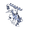
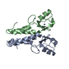
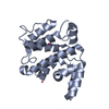
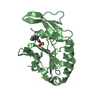
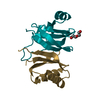
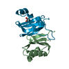
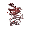
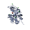
[English] 日本語
Yorodumi
- PDB-2ybf: Complex of Rad18 (Rad6 binding domain) with Rad6b -

+
Open data


ID or keywords:

Loading...

-
Basic information

Entry
Database: PDB / ID: 2ybf
TitleComplex of Rad18 (Rad6 binding domain) with Rad6b
Components
  • E3 UBIQUITIN-PROTEIN LIGASE RAD18
  • UBIQUITIN-CONJUGATING ENZYME E2 B
KeywordsLIGASE / E2 UBIQUITIN CONJUGATING ENZYME / E3 UBIQUITIN LIGASE / TRANSLESION SYNTHESIS / PCNA UBIQUITINATION
Function / homology
Function and homology information


synaptonemal complex organization / negative regulation of post-translational protein modification / positive regulation of reciprocal meiotic recombination / chiasma assembly / Rad6-Rad18 complex / positive regulation of chromosome segregation / HULC complex / sperm axoneme assembly / meiotic telomere clustering / Y-form DNA binding ...synaptonemal complex organization / negative regulation of post-translational protein modification / positive regulation of reciprocal meiotic recombination / chiasma assembly / Rad6-Rad18 complex / positive regulation of chromosome segregation / HULC complex / sperm axoneme assembly / meiotic telomere clustering / Y-form DNA binding / nuclear inclusion body / protein K11-linked ubiquitination / XY body / negative regulation of cAMP-mediated signaling / postreplication repair / E2 ubiquitin-conjugating enzyme / negative regulation of reproductive process / negative regulation of developmental process / protein monoubiquitination / ubiquitin conjugating enzyme activity / protein K63-linked ubiquitination / polyubiquitin modification-dependent protein binding / ectopic germ cell programmed cell death / protein autoubiquitination / protein K48-linked ubiquitination / response to UV / Synthesis of active ubiquitin: roles of E1 and E2 enzymes / replication fork / Recognition of DNA damage by PCNA-containing replication complex / RING-type E3 ubiquitin transferase / protein polyubiquitination / ubiquitin-protein transferase activity / ubiquitin protein ligase activity / Antigen processing: Ubiquitination & Proteasome degradation / positive regulation of canonical Wnt signaling pathway / E3 ubiquitin ligases ubiquitinate target proteins / chromatin organization / site of double-strand break / single-stranded DNA binding / ubiquitin-dependent protein catabolic process / spermatogenesis / proteasome-mediated ubiquitin-dependent protein catabolic process / in utero embryonic development / damaged DNA binding / nuclear body / protein stabilization / protein ubiquitination / response to xenobiotic stimulus / DNA repair / centrosome / apoptotic process / ubiquitin protein ligase binding / DNA damage response / protein-containing complex binding / chromatin / negative regulation of apoptotic process / nucleoplasm / ATP binding / identical protein binding / nucleus / metal ion binding / plasma membrane / cytoplasm
Similarity search - Function
E3 ubiquitin-protein ligase Rad18 / Rad18-like CCHC zinc finger / Zinc finger, C3HC4 type (RING finger) / SAP domain superfamily / SAP domain / SAP motif profile. / Putative DNA-binding (bihelical) motif predicted to be involved in chromosomal organisation / SAP domain / Ubiquitin Conjugating Enzyme / Ubiquitin Conjugating Enzyme ...E3 ubiquitin-protein ligase Rad18 / Rad18-like CCHC zinc finger / Zinc finger, C3HC4 type (RING finger) / SAP domain superfamily / SAP domain / SAP motif profile. / Putative DNA-binding (bihelical) motif predicted to be involved in chromosomal organisation / SAP domain / Ubiquitin Conjugating Enzyme / Ubiquitin Conjugating Enzyme / Rad18, zinc finger UBZ4-type / Zinc finger UBZ4-type profile. / Ubiquitin-conjugating enzyme, active site / Ubiquitin-conjugating (UBC) active site signature. / Ubiquitin-conjugating enzyme E2, catalytic domain homologues / Ubiquitin-conjugating enzyme E2 / Ubiquitin-conjugating enzyme / Ubiquitin-conjugating (UBC) core domain profile. / Ubiquitin-conjugating enzyme/RWD-like / Zinc finger, RING-type, conserved site / Zinc finger RING-type signature. / Ring finger / Zinc finger RING-type profile. / Zinc finger, RING-type / Zinc finger, RING/FYVE/PHD-type / Roll / Alpha Beta
Similarity search - Domain/homology
BETA-MERCAPTOETHANOL / Ubiquitin-conjugating enzyme E2 B / E3 ubiquitin-protein ligase RAD18
Similarity search - Component
Biological speciesHOMO SAPIENS (human)
MethodX-RAY DIFFRACTION / SYNCHROTRON / MOLECULAR REPLACEMENT / Resolution: 2 Å
AuthorsHibbert, R.G. / Sixma, T.K.
CitationJournal: Proc.Natl.Acad.Sci.USA / Year: 2011
Title: E3 Ligase Rad18 Promotes Monoubiquitination Rather Than Ubiquitin Chain Formation by E2 Enzyme Rad6.
Authors: Hibbert, R.G. / Huang, A. / Boelens, R. / Sixma, T.K.
History
DepositionMar 8, 2011Deposition site: PDBE / Processing site: PDBE
Revision 1.0Apr 20, 2011Provider: repository / Type: Initial release
Revision 1.1May 8, 2011Group: Version format compliance
Revision 1.2Jul 13, 2011Group: Version format compliance
Revision 1.3Dec 20, 2023Group: Data collection / Database references ...Data collection / Database references / Derived calculations / Other / Refinement description
Category: chem_comp_atom / chem_comp_bond ...chem_comp_atom / chem_comp_bond / database_2 / pdbx_database_status / pdbx_initial_refinement_model / pdbx_struct_conn_angle / struct_conn / struct_site
Item: _database_2.pdbx_DOI / _database_2.pdbx_database_accession ..._database_2.pdbx_DOI / _database_2.pdbx_database_accession / _pdbx_database_status.status_code_sf / _pdbx_struct_conn_angle.ptnr1_auth_seq_id / _pdbx_struct_conn_angle.ptnr1_symmetry / _pdbx_struct_conn_angle.ptnr3_auth_seq_id / _pdbx_struct_conn_angle.ptnr3_symmetry / _pdbx_struct_conn_angle.value / _struct_conn.pdbx_dist_value / _struct_conn.ptnr2_auth_seq_id / _struct_conn.ptnr2_symmetry / _struct_site.pdbx_auth_asym_id / _struct_site.pdbx_auth_comp_id / _struct_site.pdbx_auth_seq_id

-
Structure visualization

Structure viewerMolecule:
MolmilJmol/JSmol

Downloads & links

-
Assembly

Deposited unit
A: UBIQUITIN-CONJUGATING ENZYME E2 B
B: E3 UBIQUITIN-PROTEIN LIGASE RAD18
hetero molecules


Theoretical massNumber of molelcules
Total (without water)20,8396
Polymers20,5462
Non-polymers2934
Water1,53185
1


  • Idetical with deposited unit
  • defined by software
TypeNameSymmetry operationNumber
identity operation1_555x,y,z1
Buried area2470 Å2
ΔGint-47.4 kcal/mol
Surface area9470 Å2
MethodPISA
Unit cell
Length a, b, c (Å)58.210, 58.210, 167.070
Angle α, β, γ (deg.)90.00, 90.00, 120.00
Int Tables number179
Space group name H-MP6522

-
Components

-
Protein / Protein/peptide , 2 types, 2 molecules AB

#1: Protein UBIQUITIN-CONJUGATING ENZYME E2 B / RAD6 HOMOLOG B / HR6B / HHR6B / UBIQUITIN CARRIER PROTEIN B / UBIQUITIN-CONJUGATING ENZYME E2-17 ...RAD6 HOMOLOG B / HR6B / HHR6B / UBIQUITIN CARRIER PROTEIN B / UBIQUITIN-CONJUGATING ENZYME E2-17 KDA / UBIQUITIN-PROTEIN LIGASE B / RAD6B


Mass: 17328.283 Da / Num. of mol.: 1
Source method: isolated from a genetically manipulated source
Source: (gene. exp.) HOMO SAPIENS (human) / Production host: ESCHERICHIA COLI (E. coli) / Strain (production host): BL21(DE3) / References: UniProt: P63146, ubiquitin-protein ligase
#2: Protein/peptide E3 UBIQUITIN-PROTEIN LIGASE RAD18 / POSTREPLICATION REPAIR PROTEIN RAD18 / HHR18 / HRAD18 / RING FINGER PROTEIN 73


Mass: 3217.808 Da / Num. of mol.: 1 / Fragment: RAD6-BINDING DOMAIN, RESIDUES 340-366 / Source method: obtained synthetically / Source: (synth.) HOMO SAPIENS (human)
References: UniProt: Q9NS91, Ligases; Forming carbon-nitrogen bonds; Acid-amino-acid ligases (peptide synthases)

-
Non-polymers , 4 types, 89 molecules

#3: Chemical ChemComp-BME / BETA-MERCAPTOETHANOL


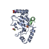
Mass: 78.133 Da / Num. of mol.: 1 / Source method: obtained synthetically / Formula: C2H6OS
#4: Chemical ChemComp-SO4 / SULFATE ION


Mass: 96.063 Da / Num. of mol.: 2 / Source method: obtained synthetically / Formula: SO4
#5: Chemical ChemComp-NA / SODIUM ION


Mass: 22.990 Da / Num. of mol.: 1 / Source method: obtained synthetically / Formula: Na
#6: Water ChemComp-HOH / water


Mass: 18.015 Da / Num. of mol.: 85 / Source method: isolated from a natural source / Formula: H2O

-
Experimental details

-
Experiment

ExperimentMethod: X-RAY DIFFRACTION / Number of used crystals: 1

-
Sample preparation

CrystalDensity Matthews: 1.99 Å3/Da / Density % sol: 38.1 % / Description: NONE
Crystal growpH: 6.5 / Details: pH 6.5

-
Data collection

DiffractionMean temperature: 100 K
Diffraction sourceSource: SYNCHROTRON / Site: ESRF / Beamline: ID23-2 / Wavelength: 0.873
DetectorType: MARMOSAIC 225 mm CCD / Detector: CCD / Date: Mar 12, 2009
RadiationProtocol: SINGLE WAVELENGTH / Monochromatic (M) / Laue (L): M / Scattering type: x-ray
Radiation wavelengthWavelength: 0.873 Å / Relative weight: 1
ReflectionResolution: 2→24.92 Å / Num. obs: 12029 / % possible obs: 99.3 % / Observed criterion σ(I): 2 / Redundancy: 12.9 % / Biso Wilson estimate: 22.7 Å2 / Rmerge(I) obs: 0.13 / Net I/σ(I): 16
Reflection shellResolution: 2→2.11 Å / Redundancy: 12.1 % / Rmerge(I) obs: 0.72 / Mean I/σ(I) obs: 4.7 / % possible all: 95.5

-
Processing

Software
NameVersionClassification
REFMAC5.5.0099refinement
XDSdata reduction
SCALAdata scaling
PHASERphasing
RefinementMethod to determine structure: MOLECULAR REPLACEMENT
Starting model: PDB ENTRY 1AYZ
Resolution: 2→24.92 Å / Cor.coef. Fo:Fc: 0.942 / Cor.coef. Fo:Fc free: 0.905 / SU B: 9.518 / SU ML: 0.124 / Cross valid method: THROUGHOUT / ESU R: 0.22 / ESU R Free: 0.181 / Stereochemistry target values: MAXIMUM LIKELIHOOD / Details: HYDROGENS HAVE BEEN ADDED IN THE RIDING POSITIONS.
RfactorNum. reflection% reflectionSelection details
Rfree0.24204 599 5 %RANDOM
Rwork0.1989 ---
obs0.20099 11364 99.35 %-
Solvent computationIon probe radii: 0.8 Å / Shrinkage radii: 0.8 Å / VDW probe radii: 1.4 Å / Solvent model: MASK
Displacement parametersBiso mean: 29.216 Å2
Baniso -1Baniso -2Baniso -3
1--0.95 Å2-0.48 Å20 Å2
2---0.95 Å20 Å2
3---1.43 Å2
Refinement stepCycle: LAST / Resolution: 2→24.92 Å
ProteinNucleic acidLigandSolventTotal
Num. atoms1390 0 15 85 1490
Refine LS restraints
Refine-IDTypeDev idealDev ideal targetNumber
X-RAY DIFFRACTIONr_bond_refined_d0.0060.0221486
X-RAY DIFFRACTIONr_bond_other_d0.0010.021056
X-RAY DIFFRACTIONr_angle_refined_deg0.9271.9622022
X-RAY DIFFRACTIONr_angle_other_deg0.7793.0012571
X-RAY DIFFRACTIONr_dihedral_angle_1_deg4.5985183
X-RAY DIFFRACTIONr_dihedral_angle_2_deg33.86424.56881
X-RAY DIFFRACTIONr_dihedral_angle_3_deg11.70515259
X-RAY DIFFRACTIONr_dihedral_angle_4_deg13.1131512
X-RAY DIFFRACTIONr_chiral_restr0.0570.2207
X-RAY DIFFRACTIONr_gen_planes_refined0.0030.0211656
X-RAY DIFFRACTIONr_gen_planes_other0.0010.02297
X-RAY DIFFRACTIONr_nbd_refined
X-RAY DIFFRACTIONr_nbd_other
X-RAY DIFFRACTIONr_nbtor_refined
X-RAY DIFFRACTIONr_nbtor_other
X-RAY DIFFRACTIONr_xyhbond_nbd_refined
X-RAY DIFFRACTIONr_xyhbond_nbd_other
X-RAY DIFFRACTIONr_metal_ion_refined
X-RAY DIFFRACTIONr_metal_ion_other
X-RAY DIFFRACTIONr_symmetry_vdw_refined
X-RAY DIFFRACTIONr_symmetry_vdw_other
X-RAY DIFFRACTIONr_symmetry_hbond_refined
X-RAY DIFFRACTIONr_symmetry_hbond_other
X-RAY DIFFRACTIONr_symmetry_metal_ion_refined
X-RAY DIFFRACTIONr_symmetry_metal_ion_other
X-RAY DIFFRACTIONr_mcbond_it0.3591.5881
X-RAY DIFFRACTIONr_mcbond_other0.0781.5340
X-RAY DIFFRACTIONr_mcangle_it0.68121440
X-RAY DIFFRACTIONr_mcangle_other
X-RAY DIFFRACTIONr_scbond_it1.0953605
X-RAY DIFFRACTIONr_scbond_other
X-RAY DIFFRACTIONr_scangle_it1.7854.5575
X-RAY DIFFRACTIONr_scangle_other
X-RAY DIFFRACTIONr_long_range_B_refined
X-RAY DIFFRACTIONr_long_range_B_other
X-RAY DIFFRACTIONr_rigid_bond_restr
X-RAY DIFFRACTIONr_sphericity_free
X-RAY DIFFRACTIONr_sphericity_bonded
LS refinement shellResolution: 2.001→2.053 Å / Total num. of bins used: 20
RfactorNum. reflection% reflection
Rfree0.252 39 -
Rwork0.23 741 -
obs--91.02 %
Refinement TLS params.

Method: refined / Refine-ID: X-RAY DIFFRACTION

IDL112)L122)L132)L222)L232)L332)S11 (Å °)S12 (Å °)S13 (Å °)S21 (Å °)S22 (Å °)S23 (Å °)S31 (Å °)S32 (Å °)S33 (Å °)T112)T122)T132)T222)T232)T332)Origin x (Å)Origin y (Å)Origin z (Å)
18.27566.5613-1.99819.58111.71024.1273-0.24230.3319-0.1754-0.26170.25250.2959-0.04940.0331-0.01020.01430.00610.00660.1123-0.01940.025234.901221.7186-7.3071
24.0694-0.1843-0.96351.60061.45353.0827-0.0710.09630.0998-0.07730.00390.1001-0.1456-0.08010.06710.0512-0.0144-0.01040.06440.0140.011423.153832.16582.5521
326.6128-4.26499.03215.8483-3.06110.2139-0.795-1.27712.2020.84160.14110.3587-1.2852-0.49830.65390.3321-0.03390.03060.1782-0.19240.351722.587546.232613.8887
410.1623-0.086-1.90346.2544-1.629910.81430.002-0.6075-0.13170.68480.0999-0.1633-0.24830.1125-0.10190.09460.0348-0.02240.1746-0.07440.060337.154730.939610.6826
Refinement TLS group
IDRefine-IDRefine TLS-IDAuth asym-IDAuth seq-ID
1X-RAY DIFFRACTION1A3 - 20
2X-RAY DIFFRACTION2A21 - 131
3X-RAY DIFFRACTION2A1153
4X-RAY DIFFRACTION3A132 - 152
5X-RAY DIFFRACTION4B341 - 361

+
About Yorodumi

-
News

-
Feb 9, 2022. New format data for meta-information of EMDB entries

New format data for meta-information of EMDB entries

  • Version 3 of the EMDB header file is now the official format.
  • The previous official version 1.9 will be removed from the archive.

Related info.:EMDB header

External links:wwPDB to switch to version 3 of the EMDB data model

-
Aug 12, 2020. Covid-19 info

Covid-19 info

URL: https://pdbjlvh1.pdbj.org/emnavi/covid19.php

New page: Covid-19 featured information page in EM Navigator.

Related info.:Covid-19 info / Mar 5, 2020. Novel coronavirus structure data

+
Mar 5, 2020. Novel coronavirus structure data

Novel coronavirus structure data

Related info.:Yorodumi Speices / Aug 12, 2020. Covid-19 info

External links:COVID-19 featured content - PDBj / Molecule of the Month (242):Coronavirus Proteases

+
Jan 31, 2019. EMDB accession codes are about to change! (news from PDBe EMDB page)

EMDB accession codes are about to change! (news from PDBe EMDB page)

  • The allocation of 4 digits for EMDB accession codes will soon come to an end. Whilst these codes will remain in use, new EMDB accession codes will include an additional digit and will expand incrementally as the available range of codes is exhausted. The current 4-digit format prefixed with “EMD-” (i.e. EMD-XXXX) will advance to a 5-digit format (i.e. EMD-XXXXX), and so on. It is currently estimated that the 4-digit codes will be depleted around Spring 2019, at which point the 5-digit format will come into force.
  • The EM Navigator/Yorodumi systems omit the EMD- prefix.

Related info.:Q: What is EMD? / ID/Accession-code notation in Yorodumi/EM Navigator

External links:EMDB Accession Codes are Changing Soon! / Contact to PDBj

+
Jul 12, 2017. Major update of PDB

Major update of PDB

  • wwPDB released updated PDB data conforming to the new PDBx/mmCIF dictionary.
  • This is a major update changing the version number from 4 to 5, and with Remediation, in which all the entries are updated.
  • In this update, many items about electron microscopy experimental information are reorganized (e.g. em_software).
  • Now, EM Navigator and Yorodumi are based on the updated data.

External links:wwPDB Remediation / Enriched Model Files Conforming to OneDep Data Standards Now Available in the PDB FTP Archive

-
Yorodumi

Thousand views of thousand structures

  • Yorodumi is a browser for structure data from EMDB, PDB, SASBDB, etc.
  • This page is also the successor to EM Navigator detail page, and also detail information page/front-end page for Omokage search.
  • The word "yorodu" (or yorozu) is an old Japanese word meaning "ten thousand". "mi" (miru) is to see.

Related info.:EMDB / PDB / SASBDB / Comparison of 3 databanks / Yorodumi Search / Aug 31, 2016. New EM Navigator & Yorodumi / Yorodumi Papers / Jmol/JSmol / Function and homology information / Changes in new EM Navigator and Yorodumi

Read more