 Movie
Movie Controller

[English] 日本語
 Yorodumi
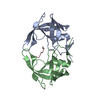
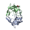
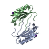
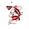
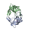
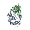
- PDB-3hvp: CONSERVED FOLDING IN RETROVIRAL PROTEASES. CRYSTAL STRUCTURE OF A... -
+ Open data

- Basic information
| Entry | Database: PDB / ID: 3hvp | ||||||
|---|---|---|---|---|---|---|---|
| Title | CONSERVED FOLDING IN RETROVIRAL PROTEASES. CRYSTAL STRUCTURE OF A SYNTHETIC HIV-1 PROTEASE | ||||||
 Components Components | UNLIGANDED HIV-1 PROTEASE | ||||||
 Keywords Keywords | HYDROLASE(ACID PROTEINASE) | ||||||
| Function / homology |  Function and homology information Function and homology informationHIV-1 retropepsin / symbiont-mediated activation of host apoptosis / retroviral ribonuclease H / exoribonuclease H / exoribonuclease H activity / DNA integration / viral genome integration into host DNA / establishment of integrated proviral latency / RNA-directed DNA polymerase / RNA stem-loop binding ...HIV-1 retropepsin / symbiont-mediated activation of host apoptosis / retroviral ribonuclease H / exoribonuclease H / exoribonuclease H activity / DNA integration / viral genome integration into host DNA / establishment of integrated proviral latency / RNA-directed DNA polymerase / RNA stem-loop binding / viral penetration into host nucleus / host multivesicular body / RNA-directed DNA polymerase activity / RNA-DNA hybrid ribonuclease activity / Transferases; Transferring phosphorus-containing groups; Nucleotidyltransferases / host cell / viral nucleocapsid / DNA recombination / DNA-directed DNA polymerase / aspartic-type endopeptidase activity / Hydrolases; Acting on ester bonds / DNA-directed DNA polymerase activity / symbiont-mediated suppression of host gene expression / viral translational frameshifting / symbiont entry into host cell / lipid binding / host cell nucleus / host cell plasma membrane / virion membrane / structural molecule activity / proteolysis / DNA binding / zinc ion binding Similarity search - Function | ||||||
| Biological species |   Human immunodeficiency virus 1 Human immunodeficiency virus 1 | ||||||
| Method |  X-RAY DIFFRACTION / Resolution: 2.8 Å X-RAY DIFFRACTION / Resolution: 2.8 Å | ||||||
 Authors Authors | Wlodawer, A. / Jaskolski, M. / Miller, M. | ||||||
 Citation Citation | Journal: Science / Year: 1989 Title: Conserved folding in retroviral proteases: crystal structure of a synthetic HIV-1 protease. Authors: Wlodawer, A. / Miller, M. / Jaskolski, M. / Sathyanarayana, B.K. / Baldwin, E. / Weber, I.T. / Selk, L.M. / Clawson, L. / Schneider, J. / Kent, S.B. #1: Journal: Science / Year: 1989Title: Molecular Modeling of the HIV-1 Protease and its Substrate Binding Site Authors: Weber, I.T. / Miller, M. / Jaskolski, M. / Leis, J. / Skalka, A.M. / Wlodawer, A. #2: Journal: Nature / Year: 1989Title: Crystal Structure of a Retroviral Protease Proves Relationship to Aspartic Protease Family Authors: Miller, M. / Jaskolski, M. / Rao, J.K.M. / Leis, J. / Wlodawer, A. #3: Journal: Cell(Cambridge,Mass.) / Year: 1988Title: Enzymatic Activity of a Synthetic 99 Residue Protein Corresponding to the Putative HIV-1 Protease Authors: Schneider, J. / Kent, S.B.H. | ||||||
| History |
| ||||||
| Remark 700 | SHEET THE DIMER INTERFACE IS COMPOSED OF INTERDIGITATED N- AND C-TERMINI FROM BOTH SUBUNITS FORMING ...SHEET THE DIMER INTERFACE IS COMPOSED OF INTERDIGITATED N- AND C-TERMINI FROM BOTH SUBUNITS FORMING A FOUR-STRANDED ANTIPARALLEL BETA-SHEET. BECAUSE OF LIMITATIONS IMPOSED BY THE PROTEIN DATA BANK FORMAT IT IS NOT POSSIBLE TO PRESENT THIS SHEET ON SHEET RECORDS. INSTEAD THIS SHEET IS SPECIFIED IN THIS REMARK. STRANDS 1 AND 3 ARE FROM THE MOLECULE IN THIS ENTRY AND STRANDS 2 AND 4 ARE FROM THE SYMMETRY RELATED MOLECULE. INT 4 PRO 1 THR 4 0 INT 4 THR 96 PHE 99 -1 INT 4 THR 96 PHE 99 -1 INT 4 PRO 1 THR 4 -1 |
- Structure visualization
| Structure viewer | Molecule: MolmilJmol/JSmol |
|---|
- Downloads & links
-Download
| PDBx/mmCIF format | 3hvp.cif.gz | 32.7 KB | Display | PDBx/mmCIF format |
|---|---|---|---|---|
| PDB format | pdb3hvp.ent.gz | 21.2 KB | Display | PDB format |
| PDBx/mmJSON format | 3hvp.json.gz | Tree view | PDBx/mmJSON format | |
| Others |  Other downloads Other downloads |
-Validation report
| Arichive directory | https://data.pdbj.org/pub/pdb/validation_reports/hv/3hvpftp://data.pdbj.org/pub/pdb/validation_reports/hv/3hvp | HTTPS FTP |
|---|
-Related structure data
| Similar structure data |
|---|


-Links
 PDBj
PDBj

- Assembly
| Deposited unit | 
| ||||||||
|---|---|---|---|---|---|---|---|---|---|
| 1 | 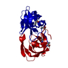
| ||||||||
| Unit cell |
| ||||||||
| Details | THE ACTIVE ENZYME IS FORMED BY TWO CRYSTALLOGRAPHICALLY RELATED MOLECULES. TO GENERATE THE OTHER MOLECULE ONE MUST APPLY THE CRYSTALLOGRAPHIC TWO-FOLD (Y, X, -Z) TO THE COORDINATES IN THIS ENTRY. |
-Components
| #1: Protein | Mass: 10764.636 Da / Num. of mol.: 1 Source method: isolated from a genetically manipulated source Source: (gene. exp.) Human immunodeficiency virus 1 / Genus: Lentivirus / Production host:  |
|---|---|
| Has protein modification | Y |
-Experimental details
-Experiment
| Experiment | Method: X-RAY DIFFRACTION |
|---|
- Sample preparation
| Crystal | Density Matthews: 3.12 Å3/Da / Density % sol: 60.59 % | ||||||||||||||||||||||||
|---|---|---|---|---|---|---|---|---|---|---|---|---|---|---|---|---|---|---|---|---|---|---|---|---|---|
| Crystal grow | *PLUS Method: vapor diffusion | ||||||||||||||||||||||||
| Components of the solutions | *PLUS
|
-Data collection
| Reflection | *PLUS Highest resolution: 2.8 Å / Num. all: 26037 / Num. obs: 3225 / Rmerge(I) obs: 1.5 |
|---|
- Processing
| Software | Name: PROFFT / Classification: refinement | ||||||||||||||||||||||||||||||||||||||||||||||||||||||||||||||||||||||||||||||||||||
|---|---|---|---|---|---|---|---|---|---|---|---|---|---|---|---|---|---|---|---|---|---|---|---|---|---|---|---|---|---|---|---|---|---|---|---|---|---|---|---|---|---|---|---|---|---|---|---|---|---|---|---|---|---|---|---|---|---|---|---|---|---|---|---|---|---|---|---|---|---|---|---|---|---|---|---|---|---|---|---|---|---|---|---|---|---|
| Refinement | Rfactor obs: 0.184 / Highest resolution: 2.8 Å | ||||||||||||||||||||||||||||||||||||||||||||||||||||||||||||||||||||||||||||||||||||
| Refinement step | Cycle: LAST / Highest resolution: 2.8 Å
| ||||||||||||||||||||||||||||||||||||||||||||||||||||||||||||||||||||||||||||||||||||
| Refine LS restraints |
| ||||||||||||||||||||||||||||||||||||||||||||||||||||||||||||||||||||||||||||||||||||
| Refinement | *PLUS Highest resolution: 2.8 Å / Rfactor obs: 0.184 | ||||||||||||||||||||||||||||||||||||||||||||||||||||||||||||||||||||||||||||||||||||
| Solvent computation | *PLUS | ||||||||||||||||||||||||||||||||||||||||||||||||||||||||||||||||||||||||||||||||||||
| Displacement parameters | *PLUS Biso mean: 16.9 Å2 |
