 Movie
Movie Controller


+ Open data

- Basic information
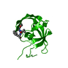
| Entry | Database: PDB / ID: 4npu | ||||||
|---|---|---|---|---|---|---|---|
| Title | Crystal Structure of HIV-1 Protease Multiple Mutant P51 | ||||||
 Components Components | Protease | ||||||
 Keywords Keywords | HYDROLASE / HIV-1 protease / P51 / multiple mutant / drug resistance | ||||||
| Function / homology |  Function and homology information Function and homology informationhost multivesicular body / aspartic-type endopeptidase activity / virion membrane / proteolysis Similarity search - Function | ||||||
| Biological species |   Human immunodeficiency virus 1 Human immunodeficiency virus 1 | ||||||
| Method |  X-RAY DIFFRACTION / SYNCHROTRON / MOLECULAR REPLACEMENT / Resolution: 1.5 Å X-RAY DIFFRACTION / SYNCHROTRON / MOLECULAR REPLACEMENT / Resolution: 1.5 Å | ||||||
 Authors Authors | Zhang, Y. / Weber, I.T. | ||||||
 Citation Citation | Journal: Acs Chem.Biol. / Year: 2014 Title: Structures of darunavir-resistant HIV-1 protease mutant reveal atypical binding of darunavir to wide open flaps. Authors: Zhang, Y. / Chang, Y.C. / Louis, J.M. / Wang, Y.F. / Harrison, R.W. / Weber, I.T. | ||||||
| History |
|
- Structure visualization
| Structure viewer | Molecule: MolmilJmol/JSmol |
|---|
- Downloads & links
-Download
| PDBx/mmCIF format | 4npu.cif.gz | 95.1 KB | Display | PDBx/mmCIF format |
|---|---|---|---|---|
| PDB format | pdb4npu.ent.gz | 73 KB | Display | PDB format |
| PDBx/mmJSON format | 4npu.json.gz | Tree view | PDBx/mmJSON format | |
| Others |  Other downloads Other downloads |
-Validation report
| Arichive directory | https://data.pdbj.org/pub/pdb/validation_reports/np/4npuftp://data.pdbj.org/pub/pdb/validation_reports/np/4npu | HTTPS FTP |
|---|
-Related structure data
| Related structure data |  4nptC 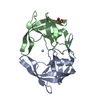 3uf3S C: citing same article ( S: Starting model for refinement |
|---|---|
| Similar structure data |


-Links
 PDBj
PDBj
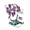
- Assembly
| Deposited unit | 
| ||||||||
|---|---|---|---|---|---|---|---|---|---|
| 1 |
| ||||||||
| Unit cell |
|
-Components
| #1: Protein | Mass: 10784.627 Da / Num. of mol.: 2 Mutation: Q7K, D25N, V32I, L33F, N37S, V54M, C67A, A82I, I84V, C95A Source method: isolated from a genetically manipulated source Source: (gene. exp.) Human immunodeficiency virus 1 / Gene: pol / Plasmid: pET11a / Production host:  #2: Water | ChemComp-HOH / |  Mass: 18.015 Da / Num. of mol.: 134 / Source method: isolated from a natural source / Formula: H2O Mass: 18.015 Da / Num. of mol.: 134 / Source method: isolated from a natural source / Formula: H2O |
|---|
-Experimental details
-Experiment
| Experiment | Method: X-RAY DIFFRACTION / Number of used crystals: 1 |
|---|
- Sample preparation
| Crystal | Density Matthews: 2.57 Å3/Da / Density % sol: 52.08 % |
|---|---|
| Crystal grow | Temperature: 298 K / Method: vapor diffusion, hanging drop / pH: 6.5 Details: 0.1 M imidazole, 1.0 M sodium acetate trihydrate, pH 6.5, VAPOR DIFFUSION, HANGING DROP, temperature 298K |
-Data collection
| Diffraction | Mean temperature: 100 K |
|---|---|
| Diffraction source | Source: SYNCHROTRON / Site: APS  / Beamline: 22-BM / Wavelength: 1 Å / Beamline: 22-BM / Wavelength: 1 Å |
| Detector | Type: MARMOSAIC 225 mm CCD / Detector: CCD / Date: Jun 29, 2012 |
| Radiation | Monochromator: SI220 CHANNEL / Protocol: SINGLE WAVELENGTH / Monochromatic (M) / Laue (L): M / Scattering type: x-ray |
| Radiation wavelength | Wavelength: 1 Å / Relative weight: 1 |
| Reflection | Resolution: 1.5→50 Å / Num. all: 34414 / Num. obs: 34414 / % possible obs: 99.4 % / Observed criterion σ(F): 0 / Observed criterion σ(I): 0 / Redundancy: 8 % / Biso Wilson estimate: 22.7 Å2 / Rmerge(I) obs: 0.067 / Net I/σ(I): 23.2 |
| Reflection shell | Resolution: 1.5→1.55 Å / Redundancy: 3.4 % / Rmerge(I) obs: 0.277 / Mean I/σ(I) obs: 4.6 / Num. unique all: 3310 / % possible all: 95.5 |
- Processing
| Software |
| |||||||||||||||||||||||||||||||||||||||||||||||||||||||||||||||||||||||||||
|---|---|---|---|---|---|---|---|---|---|---|---|---|---|---|---|---|---|---|---|---|---|---|---|---|---|---|---|---|---|---|---|---|---|---|---|---|---|---|---|---|---|---|---|---|---|---|---|---|---|---|---|---|---|---|---|---|---|---|---|---|---|---|---|---|---|---|---|---|---|---|---|---|---|---|---|---|
| Refinement | Method to determine structure: MOLECULAR REPLACEMENT Starting model: PDB entry 3UF3 Resolution: 1.5→23.64 Å / Cor.coef. Fo:Fc: 0.97 / Cor.coef. Fo:Fc free: 0.96 / SU B: 2.347 / SU ML: 0.041 / Cross valid method: THROUGHOUT / σ(F): 0 / ESU R: 0.076 / ESU R Free: 0.069 / Stereochemistry target values: MAXIMUM LIKELIHOOD / Details: HYDROGENS HAVE BEEN ADDED IN THE RIDING POSITIONS
| |||||||||||||||||||||||||||||||||||||||||||||||||||||||||||||||||||||||||||
| Solvent computation | Ion probe radii: 0.8 Å / Shrinkage radii: 0.8 Å / VDW probe radii: 1.2 Å / Solvent model: MASK | |||||||||||||||||||||||||||||||||||||||||||||||||||||||||||||||||||||||||||
| Displacement parameters | Biso mean: 25.633 Å2
| |||||||||||||||||||||||||||||||||||||||||||||||||||||||||||||||||||||||||||
| Refinement step | Cycle: LAST / Resolution: 1.5→23.64 Å
| |||||||||||||||||||||||||||||||||||||||||||||||||||||||||||||||||||||||||||
| Refine LS restraints |
| |||||||||||||||||||||||||||||||||||||||||||||||||||||||||||||||||||||||||||
| LS refinement shell | Resolution: 1.5→1.54 Å / Total num. of bins used: 20
|
