- PDB-3htn: Crystal structure of a putative dna binding protein (bt_1116) fro... -
+
Open data
ID or keywords:
Loading...
-
Basic information
Entry
Database: PDB / ID: 3htn
Title
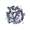
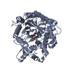
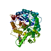
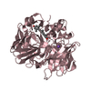
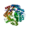
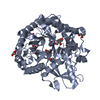
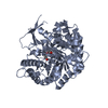
Crystal structure of a putative dna binding protein (bt_1116) from bacteroides thetaiotaomicron vpi-5482 at 1.50 A resolution
Components
Putative DNA binding protein
Keywords
METAL BINDING PROTEIN / Duf269 family protein / structural genomics / Joint Center for Structural Genomics / JCSG / Protein Structure Initiative / PSI-2
Mass: 18.015 Da / Num. of mol.: 553 / Source method: isolated from a natural source / Formula: H2O
-
Details
Has protein modification
Y
Sequence details
THE CONSTRUCT WAS EXPRESSED WITH A PURIFICATION TAG MGSDKIHHHHHHENLYFQG. THE TAG WAS REMOVED WITH ...THE CONSTRUCT WAS EXPRESSED WITH A PURIFICATION TAG MGSDKIHHHHHHENLYFQG. THE TAG WAS REMOVED WITH TEV PROTEASE LEAVING ONLY A GLYCINE (0) FOLLOWED BY THE TARGET SEQUENCE. THE CLONED CONSTRUCT CONTAINS RESIDUES 38-185 OF THE FULL LENGTH PROTEIN.
-
Experimental details
-
Experiment
Experiment
Method: X-RAY DIFFRACTION / Number of used crystals: 1
-
Sample preparation
Crystal
Density Matthews: 2.53 Å3/Da / Density % sol: 51.31 %
Crystal grow
Temperature: 277 K / pH: 8 Details: 34.0000% polyethylene glycol 400, 0.2000M lithium sulfate, 0.1M TRIS pH 8.0, NANODROP, VAPOR DIFFUSION, SITTING DROP, temperature 277K
Type: MARMOSAIC 325 mm CCD / Detector: CCD / Date: Feb 18, 2009 / Details: FLAT MIRROR (VERTICAL FOCUSING)
Radiation
Monochromator: SINGLE CRYSTAL SI(111) BENT MONOCHROMATOR (HORIZONTAL FOCUSING) Protocol: MAD / Monochromatic (M) / Laue (L): M / Scattering type: x-ray
Radiation wavelength
ID
Wavelength (Å)
Relative weight
1
0.91837
1
2
0.97908
1
3
0.9784
1
Reflection
Resolution: 1.5→29.683 Å / Num. obs: 79258 / % possible obs: 97.1 % / Observed criterion σ(I): -3 / Redundancy: 3.77 % / Biso Wilson estimate: 16.81 Å2 / Rmerge(I) obs: 0.048 / Net I/σ(I): 10.56
Reflection shell
Resolution: 1.5→1.55 Å / Rmerge(I) obs: 0.552 / Mean I/σ(I) obs: 1.6 / % possible all: 95.2
-
Phasing
Phasing
Method: MAD
-
Processing
Software
Name
Version
Classification
NB
REFMAC
5.2.0019
refinement
PHENIX
refinement
SHELX
phasing
MolProbity
3beta29
modelbuilding
XSCALE
datascaling
PDB_EXTRACT
3.006
dataextraction
XDS
datareduction
SHELXD
phasing
autoSHARP
phasing
Refinement
Method to determine structure: MAD / Resolution: 1.5→29.68 Å / Cor.coef. Fo:Fc: 0.977 / Cor.coef. Fo:Fc free: 0.969 / Occupancy max: 1 / Occupancy min: 0.2 / SU B: 2.266 / SU ML: 0.043 / TLS residual ADP flag: LIKELY RESIDUAL / Cross valid method: THROUGHOUT / σ(F): 0 / ESU R: 0.059 / ESU R Free: 0.061 Stereochemistry target values: MAXIMUM LIKELIHOOD WITH PHASES Details: 1.HYDROGENS HAVE BEEN ADDED IN THE RIDING POSITIONS. 2.ATOM RECORD CONTAINS RESIDUAL B FACTORS ONLY. 3.A MET-INHIBITION PROTOCOL WAS USED FOR SELENOMETHIONINE INCORPORATION DURING PROTEIN ...Details: 1.HYDROGENS HAVE BEEN ADDED IN THE RIDING POSITIONS. 2.ATOM RECORD CONTAINS RESIDUAL B FACTORS ONLY. 3.A MET-INHIBITION PROTOCOL WAS USED FOR SELENOMETHIONINE INCORPORATION DURING PROTEIN EXPRESSION. THE OCCUPANCY OF THE SE ATOMS IN THE MSE RESIDUES WAS REDUCED TO 0.75 TO ACCOUNT FOR THE REDUCED SCATTERING POWER DUE TO PARTIAL S-MET INCORPORATION. 4.SULPHATE ANIONS AND PEG MOLECULES FROM CRYSTALLIZATION ARE MODELED IN THE STRUCTURE, RESPECTIVELY. 5.NI AND FE METAL IONS FROM PROTEIN EXPRESSION AND PURIFICATION ARE MODELED IN THE STRUCTURE. THE PRESENCE OF NI AND FE ARE SUPPORTED BY X-RAY FLUORESCENCE, BINDING GEOMETRY AND ANOMALOUS DIFFERENCE FOURIERS ABOVE AND BELOW THE NI AND FE ABSORPTION EDGE, RESPECTIVELY.
Rfactor
Num. reflection
% reflection
Selection details
Rfree
0.167
3974
5 %
RANDOM
Rwork
0.143
-
-
-
obs
0.145
79234
99.8 %
-
Solvent computation
Ion probe radii: 0.8 Å / Shrinkage radii: 0.8 Å / VDW probe radii: 1.2 Å / Solvent model: BABINET MODEL WITH MASK
Displacement parameters
Biso mean: 21.75 Å2
Baniso -1
Baniso -2
Baniso -3
1-
0.02 Å2
0.01 Å2
0 Å2
2-
-
0.02 Å2
0 Å2
3-
-
-
-0.04 Å2
Refinement step
Cycle: LAST / Resolution: 1.5→29.68 Å
Protein
Nucleic acid
Ligand
Solvent
Total
Num. atoms
3342
0
96
553
3991
Refine LS restraints
Refine-ID
Type
Dev ideal
Dev ideal target
Number
X-RAY DIFFRACTION
r_bond_refined_d
0.013
0.022
3811
X-RAY DIFFRACTION
r_bond_other_d
0.004
0.02
2544
X-RAY DIFFRACTION
r_angle_refined_deg
1.584
1.967
5176
X-RAY DIFFRACTION
r_angle_other_deg
1.134
3
6236
X-RAY DIFFRACTION
r_dihedral_angle_1_deg
5.308
5
493
X-RAY DIFFRACTION
r_dihedral_angle_2_deg
40.897
24.463
177
X-RAY DIFFRACTION
r_dihedral_angle_3_deg
9.688
15
659
X-RAY DIFFRACTION
r_dihedral_angle_4_deg
13.132
15
20
X-RAY DIFFRACTION
r_chiral_restr
0.08
0.2
554
X-RAY DIFFRACTION
r_gen_planes_refined
0.007
0.02
4383
X-RAY DIFFRACTION
r_gen_planes_other
0.003
0.02
789
X-RAY DIFFRACTION
r_nbd_refined
0.2
0.3
643
X-RAY DIFFRACTION
r_nbd_other
0.177
0.3
2827
X-RAY DIFFRACTION
r_nbtor_refined
0.175
0.5
1869
X-RAY DIFFRACTION
r_nbtor_other
0.088
0.5
2230
X-RAY DIFFRACTION
r_xyhbond_nbd_refined
0.17
0.5
867
X-RAY DIFFRACTION
r_xyhbond_nbd_other
X-RAY DIFFRACTION
r_metal_ion_refined
0.153
0.5
2
X-RAY DIFFRACTION
r_metal_ion_other
X-RAY DIFFRACTION
r_symmetry_vdw_refined
0.211
0.3
11
X-RAY DIFFRACTION
r_symmetry_vdw_other
0.24
0.3
61
X-RAY DIFFRACTION
r_symmetry_hbond_refined
0.164
0.5
55
X-RAY DIFFRACTION
r_symmetry_hbond_other
X-RAY DIFFRACTION
r_symmetry_metal_ion_refined
X-RAY DIFFRACTION
r_symmetry_metal_ion_other
X-RAY DIFFRACTION
r_mcbond_it
1.702
3
2491
X-RAY DIFFRACTION
r_mcbond_other
0.418
3
951
X-RAY DIFFRACTION
r_mcangle_it
2.354
5
3766
X-RAY DIFFRACTION
r_scbond_it
3.678
7
1618
X-RAY DIFFRACTION
r_scangle_it
5.051
9
1410
X-RAY DIFFRACTION
r_rigid_bond_restr
X-RAY DIFFRACTION
r_sphericity_free
X-RAY DIFFRACTION
r_sphericity_bonded
LS refinement shell
Resolution: 1.5→1.54 Å / Total num. of bins used: 20
Rfactor
Num. reflection
% reflection
Rfree
0.235
309
-
Rwork
0.219
5515
-
obs
-
-
98.85 %
Refinement TLS params.
Method: refined / Refine-ID: X-RAY DIFFRACTION
ID
L11 (°2)
L12 (°2)
L13 (°2)
L22 (°2)
L23 (°2)
L33 (°2)
S11 (Å °)
S12 (Å °)
S13 (Å °)
S21 (Å °)
S22 (Å °)
S23 (Å °)
S31 (Å °)
S32 (Å °)
S33 (Å °)
T11 (Å2)
T12 (Å2)
T13 (Å2)
T22 (Å2)
T23 (Å2)
T33 (Å2)
Origin x (Å)
Origin y (Å)
Origin z (Å)
1
0.6651
-0.116
0.3944
0.4066
-0.3913
0.6363
0.063
0.1431
-0.0207
-0.0942
-0.1062
-0.1167
0.1455
0.1821
0.0432
0.0142
0.0464
0.0115
-0.0051
0.0062
-0.0228
14.8005
29.902
1.0218
2
0.9358
-0.5208
0.0602
0.6127
-0.2007
0.2298
0.0109
-0.0879
-0.0706
0.0992
-0.0283
-0.0347
0.0671
0.0257
0.0174
0.0255
0.0116
-0.0189
-0.0366
0.0149
-0.0177
11.9268
22.2079
21.8894
3
0.3396
-0.097
-0.0165
0.5234
-0.4114
0.4251
0.0313
0.0351
-0.0545
-0.0567
-0.0024
0.1057
0.0601
-0.0813
-0.0289
0.0029
-0.0204
-0.0064
-0.0314
-0.0229
-0.0072
-5.7822
26.9582
9.4922
Refinement TLS group
ID
Refine-ID
Refine TLS-ID
Auth asym-ID
Auth seq-ID
1
X-RAY DIFFRACTION
1
A
43 - 185
2
X-RAY DIFFRACTION
2
B
43 - 185
3
X-RAY DIFFRACTION
3
C
43 - 185
+
About Yorodumi
-
News
-
Feb 9, 2022. New format data for meta-information of EMDB entries
New format data for meta-information of EMDB entries
Version 3 of the EMDB header file is now the official format.
The previous official version 1.9 will be removed from the archive.
In the structure databanks used in Yorodumi, some data are registered as the other names, "COVID-19 virus" and "2019-nCoV". Here are the details of the virus and the list of structure data.
Jan 31, 2019. EMDB accession codes are about to change! (news from PDBe EMDB page)
EMDB accession codes are about to change! (news from PDBe EMDB page)
The allocation of 4 digits for EMDB accession codes will soon come to an end. Whilst these codes will remain in use, new EMDB accession codes will include an additional digit and will expand incrementally as the available range of codes is exhausted. The current 4-digit format prefixed with “EMD-” (i.e. EMD-XXXX) will advance to a 5-digit format (i.e. EMD-XXXXX), and so on. It is currently estimated that the 4-digit codes will be depleted around Spring 2019, at which point the 5-digit format will come into force.
The EM Navigator/Yorodumi systems omit the EMD- prefix.
Related info.:Q: What is EMD? / ID/Accession-code notation in Yorodumi/EM Navigator
Yorodumi is a browser for structure data from EMDB, PDB, SASBDB, etc.
This page is also the successor to EM Navigator detail page, and also detail information page/front-end page for Omokage search.
The word "yorodu" (or yorozu) is an old Japanese word meaning "ten thousand". "mi" (miru) is to see.
Related info.:EMDB / PDB / SASBDB / Comparison of 3 databanks / Yorodumi Search / Aug 31, 2016. New EM Navigator & Yorodumi / Yorodumi Papers / Jmol/JSmol / Function and homology information / Changes in new EM Navigator and Yorodumi
 Movie
Movie Controller

 Yorodumi
 Open data
 Basic information
 Components
 Keywords
 Function and homology information
 Bacteroides thetaiotaomicron VPI-5482 (bacteria)
 X-RAY DIFFRACTION /
  Authors
 Citation
 Structure visualization
 Downloads & links
 Other downloads
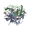


 PDBj
PDBj Assembly


 Mass: 58.693 Da / Num. of mol.: 2 / Source method: obtained synthetically / Formula: Ni
 Mass: 96.063 Da / Num. of mol.: 8 / Source method: obtained synthetically / Formula: SO4
 Mass: 238.278 Da / Num. of mol.: 5 / Source method: obtained synthetically / Formula: C10H22O6 / Comment: precipitant*YM
 Mass: 55.845 Da / Num. of mol.: 1 / Source method: obtained synthetically / Formula: Fe
 Sample preparation
 / Beamline: BL11-1 / Wavelength: 0.91837,0.97908,0.97840
 Processing