[English] 日本語
Yorodumi
- PDB-3hro: Crystal structure of a C-terminal coiled coil domain of Transient... -

+
Open data


ID or keywords:

Loading...

-
Basic information

Entry
Database: PDB / ID: 3hro
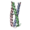
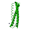
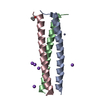
TitleCrystal structure of a C-terminal coiled coil domain of Transient receptor potential (TRP) channel subfamily P member 2 (TRPP2, polycystic kidney disease 2)
ComponentsTransient receptor potential (TRP) channel subfamily P member 2 (TRPP2), also called Polycystin-2 or polycystic kidney disease 2(PKD2)
KeywordsTRANSPORT PROTEIN / coiled coil / helix bundle / trimer / Calcium / Disease mutation / Glycoprotein / Ion transport / Ionic channel / Membrane / Phosphoprotein / Polymorphism / Transmembrane / Transport
Function / homology
Function and homology information


detection of nodal flow / metanephric smooth muscle tissue development / metanephric cortex development / metanephric cortical collecting duct development / metanephric distal tubule development / polycystin complex / mesonephric tubule development / mesonephric duct development / metanephric part of ureteric bud development / renal tubule morphogenesis ...detection of nodal flow / metanephric smooth muscle tissue development / metanephric cortex development / metanephric cortical collecting duct development / metanephric distal tubule development / polycystin complex / mesonephric tubule development / mesonephric duct development / metanephric part of ureteric bud development / renal tubule morphogenesis / determination of liver left/right asymmetry / HLH domain binding / metanephric ascending thin limb development / metanephric mesenchyme development / metanephric S-shaped body morphogenesis / basal cortex / renal artery morphogenesis / positive regulation of inositol 1,4,5-trisphosphate-sensitive calcium-release channel activity / calcium-induced calcium release activity / migrasome / cilium organization / VxPx cargo-targeting to cilium / detection of mechanical stimulus / muscle alpha-actinin binding / regulation of calcium ion import / voltage-gated monoatomic ion channel activity / placenta blood vessel development / cellular response to hydrostatic pressure / cation channel complex / cellular response to fluid shear stress / outward rectifier potassium channel activity / actinin binding / cellular response to osmotic stress / non-motile cilium / determination of left/right symmetry / inorganic cation transmembrane transport / voltage-gated monoatomic cation channel activity / aorta development / neural tube development / motile cilium / voltage-gated sodium channel activity / ciliary membrane / branching involved in ureteric bud morphogenesis / protein heterotetramerization / negative regulation of G1/S transition of mitotic cell cycle / spinal cord development / cytoplasmic side of endoplasmic reticulum membrane / heart looping / centrosome duplication / voltage-gated potassium channel activity / cell surface receptor signaling pathway via JAK-STAT / potassium channel activity / embryonic placenta development / voltage-gated calcium channel activity / transcription regulator inhibitor activity / monoatomic cation channel activity / cytoskeletal protein binding / cellular response to cAMP / release of sequestered calcium ion into cytosol / potassium ion transmembrane transport / sodium ion transmembrane transport / cytoplasmic vesicle membrane / cellular response to calcium ion / liver development / basal plasma membrane / lumenal side of endoplasmic reticulum membrane / cellular response to reactive oxygen species / establishment of localization in cell / phosphoprotein binding / protein tetramerization / calcium ion transmembrane transport / Wnt signaling pathway / intracellular calcium ion homeostasis / calcium ion transport / mitotic spindle / positive regulation of nitric oxide biosynthetic process / cell-cell junction / lamellipodium / regulation of cell population proliferation / heart development / ATPase binding / positive regulation of cytosolic calcium ion concentration / basolateral plasma membrane / protein homotetramerization / transmembrane transporter binding / cell surface receptor signaling pathway / regulation of cell cycle / ciliary basal body / cilium / signaling receptor binding / negative regulation of cell population proliferation / calcium ion binding / positive regulation of gene expression / endoplasmic reticulum membrane / endoplasmic reticulum / Golgi apparatus / protein homodimerization activity / positive regulation of transcription by RNA polymerase II / extracellular exosome / identical protein binding
Similarity search - Function
Ferredoxin I 4Fe-4S cluster domain / : / Polycystic kidney disease type 2 protein / Polycystin domain / Polycystin domain / Polycystin cation channel, PKD1/PKD2 / Polycystin cation channel / Voltage-dependent channel domain superfamily / EF-hand calcium-binding domain profile. / EF-hand domain / EF-hand domain pair
Similarity search - Domain/homology
Biological speciesHomo sapiens (human)
MethodX-RAY DIFFRACTION / SYNCHROTRON / MOLECULAR REPLACEMENT / molecular replacement / Resolution: 1.9 Å
AuthorsYu, Y. / Ulbrich, M.H. / Li, M.-H. / Buraei, Z. / Chen, X.-Z. / Ong, A.C.M. / Tong, L. / Isacoff, E.Y. / Yang, J.
CitationJournal: Proc.Natl.Acad.Sci.USA / Year: 2009
Title: Structural and molecular basis of the assembly of the TRPP2/PKD1 complex.
Authors: Yu, Y. / Ulbrich, M.H. / Li, M.H. / Buraei, Z. / Chen, X.Z. / Ong, A.C. / Tong, L. / Isacoff, E.Y. / Yang, J.
History
DepositionJun 9, 2009Deposition site: RCSB / Processing site: RCSB
Revision 1.0Jul 28, 2009Provider: repository / Type: Initial release
Revision 1.1Jul 13, 2011Group: Version format compliance
Revision 1.2Nov 1, 2017Group: Refinement description / Category: software
Revision 1.3Sep 6, 2023Group: Data collection / Database references / Refinement description
Category: chem_comp_atom / chem_comp_bond ...chem_comp_atom / chem_comp_bond / database_2 / pdbx_initial_refinement_model / struct_ref_seq_dif
Item: _database_2.pdbx_DOI / _database_2.pdbx_database_accession / _struct_ref_seq_dif.details

-
Structure visualization

Structure viewerMolecule:
MolmilJmol/JSmol

Downloads & links

-
Assembly

Deposited unit
A: Transient receptor potential (TRP) channel subfamily P member 2 (TRPP2), also called Polycystin-2 or polycystic kidney disease 2(PKD2)


Theoretical massNumber of molelcules
Total (without water)5,0531
Polymers5,0531
Non-polymers00
Water57632
1
A: Transient receptor potential (TRP) channel subfamily P member 2 (TRPP2), also called Polycystin-2 or polycystic kidney disease 2(PKD2)

A: Transient receptor potential (TRP) channel subfamily P member 2 (TRPP2), also called Polycystin-2 or polycystic kidney disease 2(PKD2)

A: Transient receptor potential (TRP) channel subfamily P member 2 (TRPP2), also called Polycystin-2 or polycystic kidney disease 2(PKD2)


Theoretical massNumber of molelcules
Total (without water)15,1593
Polymers15,1593
Non-polymers00
Water543
TypeNameSymmetry operationNumber
identity operation1_555x,y,z1
crystal symmetry operation2_665-y+1,x-y+1,z1
crystal symmetry operation3_565-x+y,-x+1,z1
Buried area4120 Å2
ΔGint-49 kcal/mol
Surface area6790 Å2
MethodPISA
Unit cell
Length a, b, c (Å)32.388, 32.388, 70.300
Angle α, β, γ (deg.)90.000, 90.000, 120.000
Int Tables number173
Space group name H-MP63

-
Components

#1: Protein/peptide Transient receptor potential (TRP) channel subfamily P member 2 (TRPP2), also called Polycystin-2 or polycystic kidney disease 2(PKD2) / Polycystin-2 / Polycystic kidney disease 2 protein / Autosomal dominant polycystic kidney disease ...Polycystin-2 / Polycystic kidney disease 2 protein / Autosomal dominant polycystic kidney disease type II protein / Polycystwin / R48321


Mass: 5052.893 Da / Num. of mol.: 1
Fragment: C-terminal of Coiled Coil Domain, UNP residues 833-872
Source method: isolated from a genetically manipulated source
Source: (gene. exp.) Homo sapiens (human) / Gene: PKD2 / Plasmid: pET-28a(+) / Production host: Escherichia coli (E. coli) / Strain (production host): Rosetta 2(DE3) / References: UniProt: Q13563
#2: Water ChemComp-HOH / water


Mass: 18.015 Da / Num. of mol.: 32 / Source method: isolated from a natural source / Formula: H2O

-
Experimental details

-
Experiment

ExperimentMethod: X-RAY DIFFRACTION / Number of used crystals: 1

-
Sample preparation

CrystalDensity Matthews: 2.11 Å3/Da / Density % sol: 41.61 %
Crystal growTemperature: 293 K / Method: vapor diffusion, hanging drop / pH: 4
Details: PEG 6000, LiCl, Citric Acid, pH 4.0, vapor diffusion, hanging drop, temperature 293K

-
Data collection

DiffractionMean temperature: 100 K
Diffraction sourceSource: SYNCHROTRON / Site: NSLS / Beamline: X4C / Wavelength: 0.97927 Å
DetectorType: MAR CCD 165 mm / Detector: CCD / Date: Mar 16, 2006
RadiationMonochromator: Si 111 CHANNEL / Protocol: SINGLE WAVELENGTH / Monochromatic (M) / Laue (L): M / Scattering type: x-ray
Radiation wavelengthWavelength: 0.97927 Å / Relative weight: 1
ReflectionResolution: 1.9→30 Å / Num. obs: 3273 / % possible obs: 98.7 % / Observed criterion σ(I): -3 / Redundancy: 5.7 % / Rmerge(I) obs: 0.052 / Χ2: 1.124 / Net I/σ(I): 32.805
Reflection shellResolution: 1.9→1.97 Å / Redundancy: 3.4 % / Rmerge(I) obs: 0.228 / Mean I/σ(I) obs: 5.1 / Num. unique all: 290 / Χ2: 1.226 / % possible all: 92.9

-
Phasing

PhasingMethod: molecular replacement

-
Processing

Software
NameVersionClassificationNB
DENZOdata reduction
SCALEPACKdata scaling
PHASERphasing
CNSrefinement
PDB_EXTRACT3.005data extraction
ADSCQuantumdata collection
RefinementMethod to determine structure: MOLECULAR REPLACEMENT
Starting model: PDB ENTRY 1ZIJ
Resolution: 1.9→30 Å / Occupancy max: 1 / Occupancy min: 1 / FOM work R set: 0.872 / Cross valid method: THROUGHOUT / σ(F): 2 / Stereochemistry target values: Engh & Huber
RfactorNum. reflection% reflectionSelection details
Rfree0.227 281 8.5 %RANDOM
Rwork0.194 ---
all0.217 3190 --
obs0.197 2991 90 %-
Solvent computationBsol: 56.794 Å2
Displacement parametersBiso max: 52.47 Å2 / Biso mean: 26.404 Å2 / Biso min: 14.36 Å2
Baniso -1Baniso -2Baniso -3
1-5.439 Å20 Å20 Å2
2--5.439 Å20 Å2
3----10.877 Å2
Refinement stepCycle: LAST / Resolution: 1.9→30 Å
ProteinNucleic acidLigandSolventTotal
Num. atoms299 0 0 32 331
Refine LS restraints
Refine-IDTypeDev idealDev ideal target
X-RAY DIFFRACTIONc_bond_d0.012
X-RAY DIFFRACTIONc_angle_d1.227
X-RAY DIFFRACTIONc_mcbond_it1.4861.5
X-RAY DIFFRACTIONc_scbond_it3.0792
X-RAY DIFFRACTIONc_mcangle_it2.1312
X-RAY DIFFRACTIONc_scangle_it4.3312.5
Xplor file
Refine-IDSerial noParam fileTopol file
X-RAY DIFFRACTION1CNS_TOPPAR:protein_rep.paramCNS_TOPPAR:protein.top
X-RAY DIFFRACTION2CNS_TOPPAR:dna-rna_rep.paramCNS_TOPPAR:dna-rna.top
X-RAY DIFFRACTION3CNS_TOPPAR:water_rep.paramCNS_TOPPAR:water.top
X-RAY DIFFRACTION4CNS_TOPPAR:ion.paramCNS_TOPPAR:ion.top

+
About Yorodumi

-
News

-
Feb 9, 2022. New format data for meta-information of EMDB entries

New format data for meta-information of EMDB entries

  • Version 3 of the EMDB header file is now the official format.
  • The previous official version 1.9 will be removed from the archive.

Related info.:EMDB header

External links:wwPDB to switch to version 3 of the EMDB data model

-
Aug 12, 2020. Covid-19 info

Covid-19 info

URL: https://pdbj.org/emnavi/covid19.php

New page: Covid-19 featured information page in EM Navigator.

Related info.:Covid-19 info / Mar 5, 2020. Novel coronavirus structure data

+
Mar 5, 2020. Novel coronavirus structure data

Novel coronavirus structure data

Related info.:Yorodumi Speices / Aug 12, 2020. Covid-19 info

External links:COVID-19 featured content - PDBj / Molecule of the Month (242):Coronavirus Proteases

+
Jan 31, 2019. EMDB accession codes are about to change! (news from PDBe EMDB page)

EMDB accession codes are about to change! (news from PDBe EMDB page)

  • The allocation of 4 digits for EMDB accession codes will soon come to an end. Whilst these codes will remain in use, new EMDB accession codes will include an additional digit and will expand incrementally as the available range of codes is exhausted. The current 4-digit format prefixed with “EMD-” (i.e. EMD-XXXX) will advance to a 5-digit format (i.e. EMD-XXXXX), and so on. It is currently estimated that the 4-digit codes will be depleted around Spring 2019, at which point the 5-digit format will come into force.
  • The EM Navigator/Yorodumi systems omit the EMD- prefix.

Related info.:Q: What is EMD? / ID/Accession-code notation in Yorodumi/EM Navigator

External links:EMDB Accession Codes are Changing Soon! / Contact to PDBj

+
Jul 12, 2017. Major update of PDB

Major update of PDB

  • wwPDB released updated PDB data conforming to the new PDBx/mmCIF dictionary.
  • This is a major update changing the version number from 4 to 5, and with Remediation, in which all the entries are updated.
  • In this update, many items about electron microscopy experimental information are reorganized (e.g. em_software).
  • Now, EM Navigator and Yorodumi are based on the updated data.

External links:wwPDB Remediation / Enriched Model Files Conforming to OneDep Data Standards Now Available in the PDB FTP Archive

-
Yorodumi

Thousand views of thousand structures

  • Yorodumi is a browser for structure data from EMDB, PDB, SASBDB, etc.
  • This page is also the successor to EM Navigator detail page, and also detail information page/front-end page for Omokage search.
  • The word "yorodu" (or yorozu) is an old Japanese word meaning "ten thousand". "mi" (miru) is to see.

Related info.:EMDB / PDB / SASBDB / Comparison of 3 databanks / Yorodumi Search / Aug 31, 2016. New EM Navigator & Yorodumi / Yorodumi Papers / Jmol/JSmol / Function and homology information / Changes in new EM Navigator and Yorodumi

Read more