 Movie
Movie Controller

[English] 日本語
 Yorodumi
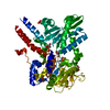
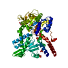
- PDB-3hqg: Crystal structure of restriction endonuclease EcoRII catalytic C-... -
+ Open data

- Basic information
| Entry | Database: PDB / ID: 3hqg | ||||||
|---|---|---|---|---|---|---|---|
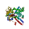
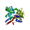
| Title | Crystal structure of restriction endonuclease EcoRII catalytic C-terminal domain in complex with cognate DNA | ||||||
 Components Components |
| ||||||
 Keywords Keywords | HYDROLASE/DNA / restriction endonuclease / EcoRII / nucleotide flipping / protein-DNA complex / DNA recognition / Endonuclease / Hydrolase / Magnesium / Nuclease / Restriction system / HYDROLASE-DNA COMPLEX | ||||||
| Function / homology |  Function and homology information Function and homology informationtype II site-specific deoxyribonuclease / type II site-specific deoxyribonuclease activity / DNA restriction-modification system / DNA binding Similarity search - Function | ||||||
| Biological species |  | ||||||
| Method |  X-RAY DIFFRACTION / SYNCHROTRON / MOLECULAR REPLACEMENT / molecular replacement / Resolution: 2.6 Å X-RAY DIFFRACTION / SYNCHROTRON / MOLECULAR REPLACEMENT / molecular replacement / Resolution: 2.6 Å | ||||||
 Authors Authors | Golovenko, D. / Manakova, E. / Grazulis, S. / Tamulaitiene, G. / Siksnys, V. | ||||||
 Citation Citation | Journal: Nucleic Acids Res. / Year: 2009 Title: Structural mechanisms for the 5'-CCWGG sequence recognition by the N- and C-terminal domains of EcoRII. Authors: Golovenko, D. / Manakova, E. / Tamulaitiene, G. / Grazulis, S. / Siksnys, V. #1: Journal: J.Mol.Biol. / Year: 2004Title: Crystal structure of type iie restriction endonuclease EcoRII reveals an autoinhibition mechanism by a novel effector-binding fold Authors: Zhou, X.E. / Wang, Y. / Reuter, M. / Mucke, M. / Kruger, D.H. / Meehan, E.J. / Chen, L. | ||||||
| History |
|
- Structure visualization
| Structure viewer | Molecule: MolmilJmol/JSmol |
|---|
- Downloads & links
-Download
| PDBx/mmCIF format | 3hqg.cif.gz | 76 KB | Display | PDBx/mmCIF format |
|---|---|---|---|---|
| PDB format | pdb3hqg.ent.gz | 52.7 KB | Display | PDB format |
| PDBx/mmJSON format | 3hqg.json.gz | Tree view | PDBx/mmJSON format | |
| Others |  Other downloads Other downloads |
-Validation report
| Arichive directory | https://data.pdbj.org/pub/pdb/validation_reports/hq/3hqgftp://data.pdbj.org/pub/pdb/validation_reports/hq/3hqg | HTTPS FTP |
|---|
-Related structure data
| Related structure data | 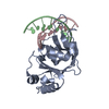 3hqfC 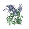 1na6S S: Starting model for refinement C: citing same article ( |
|---|---|
| Similar structure data |


-Links
 PDBj
PDBj


- Assembly
| Deposited unit | 
| ||||||||
|---|---|---|---|---|---|---|---|---|---|
| 1 | 
| ||||||||
| Unit cell |
| ||||||||
| Details | The second part of the biological assembly is generated by the two fold axis: -x+1, -y, z. |
-Components
| #1: Protein | Mass: 25577.191 Da / Num. of mol.: 1 Fragment: C-terminal catalytic domain (UNP residues 183-404) Source method: isolated from a genetically manipulated source Source: (gene. exp.) References: UniProt: P14633, type II site-specific deoxyribonuclease |
|---|---|
| #2: DNA chain | Mass: 3647.393 Da / Num. of mol.: 1 / Source method: obtained synthetically |
| #3: DNA chain | Mass: 3678.403 Da / Num. of mol.: 1 / Source method: obtained synthetically |
| #4: Chemical | ChemComp-GOL /   Mass: 92.094 Da / Num. of mol.: 1 / Source method: obtained synthetically / Formula: C3H8O3 Mass: 92.094 Da / Num. of mol.: 1 / Source method: obtained synthetically / Formula: C3H8O3 |
| #5: Water | ChemComp-HOH /  Mass: 18.015 Da / Num. of mol.: 67 / Source method: isolated from a natural source / Formula: H2O Mass: 18.015 Da / Num. of mol.: 67 / Source method: isolated from a natural source / Formula: H2O |
-Experimental details
-Experiment
| Experiment | Method: X-RAY DIFFRACTION / Number of used crystals: 1 |
|---|
- Sample preparation
| Crystal | Density Matthews: 2.07 Å3/Da / Density % sol: 40.6 % |
|---|---|
| Crystal grow | Temperature: 291 K / Method: vapor diffusion, sitting drop / pH: 8 Details: 26% PEG1500, 25% glycerol, pH 8.0, VAPOR DIFFUSION, SITTING DROP, temperature 291K |
-Data collection
| Diffraction | Mean temperature: 100 K | ||||||||||||||||||||||||||||||||||||||||||||||||||||||||||||||||||||||||||||||||||||||||
|---|---|---|---|---|---|---|---|---|---|---|---|---|---|---|---|---|---|---|---|---|---|---|---|---|---|---|---|---|---|---|---|---|---|---|---|---|---|---|---|---|---|---|---|---|---|---|---|---|---|---|---|---|---|---|---|---|---|---|---|---|---|---|---|---|---|---|---|---|---|---|---|---|---|---|---|---|---|---|---|---|---|---|---|---|---|---|---|---|---|
| Diffraction source | Source: SYNCHROTRON / Site: EMBL/DESY, HAMBURG  / Beamline: X13 / Wavelength: 0.808 Å / Beamline: X13 / Wavelength: 0.808 Å | ||||||||||||||||||||||||||||||||||||||||||||||||||||||||||||||||||||||||||||||||||||||||
| Detector | Type: MAR CCD 165 mm / Detector: CCD / Date: Nov 13, 2006 | ||||||||||||||||||||||||||||||||||||||||||||||||||||||||||||||||||||||||||||||||||||||||
| Radiation | Monochromator: Si (111), horizontally focussing / Protocol: SINGLE WAVELENGTH / Monochromatic (M) / Laue (L): M / Scattering type: x-ray | ||||||||||||||||||||||||||||||||||||||||||||||||||||||||||||||||||||||||||||||||||||||||
| Radiation wavelength | Wavelength: 0.808 Å / Relative weight: 1 | ||||||||||||||||||||||||||||||||||||||||||||||||||||||||||||||||||||||||||||||||||||||||
| Reflection | Resolution: 2.11→47.836 Å / Num. obs: 8835 / % possible obs: 99.8 % / Observed criterion σ(F): 0 / Observed criterion σ(I): 0 / Redundancy: 11.7 % / Biso Wilson estimate: 72 Å2 / Rmerge(I) obs: 0.047 / Rsym value: 0.047 / Net I/σ(I): 12.41 | ||||||||||||||||||||||||||||||||||||||||||||||||||||||||||||||||||||||||||||||||||||||||
| Reflection shell | Diffraction-ID: 1
|
-Phasing
| Phasing | Method: molecular replacement | |||||||||
|---|---|---|---|---|---|---|---|---|---|---|
| Phasing MR | Rfactor: 0.465 / Cor.coef. Fo:Fc: 0.409 / Cor.coef. Io to Ic: 0.411
|
- Processing
| Software |
| ||||||||||||||||||||||||||||||||||||||||||||||||||||||||||||||||||||||||||||||||||||||||||||||||||||
|---|---|---|---|---|---|---|---|---|---|---|---|---|---|---|---|---|---|---|---|---|---|---|---|---|---|---|---|---|---|---|---|---|---|---|---|---|---|---|---|---|---|---|---|---|---|---|---|---|---|---|---|---|---|---|---|---|---|---|---|---|---|---|---|---|---|---|---|---|---|---|---|---|---|---|---|---|---|---|---|---|---|---|---|---|---|---|---|---|---|---|---|---|---|---|---|---|---|---|---|---|---|
| Refinement | Method to determine structure: MOLECULAR REPLACEMENT Starting model: PDB ENTRY 1NA6 Resolution: 2.6→47.84 Å / Cor.coef. Fo:Fc: 0.92 / Cor.coef. Fo:Fc free: 0.875 / WRfactor Rfree: 0.29 / WRfactor Rwork: 0.223 / Occupancy max: 1 / Occupancy min: 0.01 / FOM work R set: 0.766 / SU B: 35.862 / SU ML: 0.346 / SU R Cruickshank DPI: 0.365 / SU Rfree: 0.43 / TLS residual ADP flag: LIKELY RESIDUAL / Cross valid method: THROUGHOUT / σ(F): 0 / ESU R Free: 0.43 / Stereochemistry target values: MAXIMUM LIKELIHOOD Details: HYDROGENS HAVE BEEN ADDED IN THE RIDING POSITIONS U VALUES : RESIDUAL ONLY
| ||||||||||||||||||||||||||||||||||||||||||||||||||||||||||||||||||||||||||||||||||||||||||||||||||||
| Solvent computation | Ion probe radii: 0.8 Å / Shrinkage radii: 0.8 Å / VDW probe radii: 1.4 Å / Solvent model: MASK | ||||||||||||||||||||||||||||||||||||||||||||||||||||||||||||||||||||||||||||||||||||||||||||||||||||
| Displacement parameters | Biso max: 90.36 Å2 / Biso mean: 45.712 Å2 / Biso min: 5.97 Å2
| ||||||||||||||||||||||||||||||||||||||||||||||||||||||||||||||||||||||||||||||||||||||||||||||||||||
| Refinement step | Cycle: LAST / Resolution: 2.6→47.84 Å
| ||||||||||||||||||||||||||||||||||||||||||||||||||||||||||||||||||||||||||||||||||||||||||||||||||||
| Refine LS restraints |
| ||||||||||||||||||||||||||||||||||||||||||||||||||||||||||||||||||||||||||||||||||||||||||||||||||||
| LS refinement shell | Resolution: 2.6→2.667 Å / Total num. of bins used: 20
| ||||||||||||||||||||||||||||||||||||||||||||||||||||||||||||||||||||||||||||||||||||||||||||||||||||
| Refinement TLS params. | Method: refined / Refine-ID: X-RAY DIFFRACTION
| ||||||||||||||||||||||||||||||||||||||||||||||||||||||||||||||||||||||||||||||||||||||||||||||||||||
| Refinement TLS group |
|
