 Movie
Movie Controller

[English] 日本語
 Yorodumi
- PDB-3ho1: Crystal structure of T. thermophilus Argonaute N546 mutant protei... -
+ Open data

- Basic information
| Entry | Database: PDB / ID: 3ho1 | ||||||
|---|---|---|---|---|---|---|---|
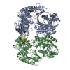
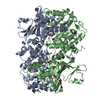
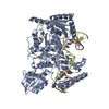
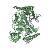
| Title | Crystal structure of T. thermophilus Argonaute N546 mutant protein complexed with DNA guide strand and 12-nt RNA target strand | ||||||
 Components Components |
| ||||||
 Keywords Keywords | NUCLEIC ACID BINDING PROTEIN/DNA/RNA / argonaute / protein-DNA-RNA complex / NUCLEIC ACID BINDING PROTEIN-DNA-RNA COMPLEX | ||||||
| Function / homology |  Function and homology information Function and homology informationHydrolases; Acting on ester bonds; Site specific endodeoxyribonucleases: cleavage is not sequence specific (deleted sub-subclass) / clearance of foreign intracellular DNA / DNA endonuclease activity / manganese ion binding / DNA replication / hydrolase activity / DNA binding / RNA binding Similarity search - Function | ||||||
| Biological species |   Thermus thermophilus (bacteria) Thermus thermophilus (bacteria) | ||||||
| Method |  X-RAY DIFFRACTION / SYNCHROTRON / MOLECULAR REPLACEMENT / Resolution: 2.6 Å X-RAY DIFFRACTION / SYNCHROTRON / MOLECULAR REPLACEMENT / Resolution: 2.6 Å | ||||||
 Authors Authors | Wang, Y. / Li, H. / Sheng, G. / Patel, D.J. | ||||||
 Citation Citation | Journal: Nature / Year: 2009 Title: Nucleation, propagation and cleavage of target RNAs in Ago silencing complexes. Authors: Wang, Y. / Juranek, S. / Li, H. / Sheng, G. / Wardle, G.S. / Tuschl, T. / Patel, D.J. | ||||||
| History |
|
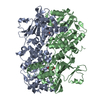
- Structure visualization
| Structure viewer | Molecule: MolmilJmol/JSmol |
|---|
- Downloads & links
-Download
| PDBx/mmCIF format | 3ho1.cif.gz | 162.2 KB | Display | PDBx/mmCIF format |
|---|---|---|---|---|
| PDB format | pdb3ho1.ent.gz | 121.2 KB | Display | PDB format |
| PDBx/mmJSON format | 3ho1.json.gz | Tree view | PDBx/mmJSON format | |
| Others |  Other downloads Other downloads |
-Validation report
| Arichive directory | https://data.pdbj.org/pub/pdb/validation_reports/ho/3ho1ftp://data.pdbj.org/pub/pdb/validation_reports/ho/3ho1 | HTTPS FTP |
|---|
-Related structure data
| Related structure data | 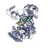 3hjfC 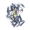 3hk2C 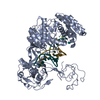 3hm9C  3hvrC 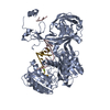 3hxmC 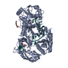 3dlhS  3hge C: citing same article ( S: Starting model for refinement |
|---|---|
| Similar structure data |


-Links
 PDBj
PDBj


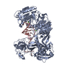
- Assembly
| Deposited unit | 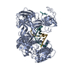
| ||||||||
|---|---|---|---|---|---|---|---|---|---|
| 1 |
| ||||||||
| Unit cell |
|
-Components
| #1: Protein | Mass: 76727.750 Da / Num. of mol.: 1 / Mutation: D546N Source method: isolated from a genetically manipulated source Source: (gene. exp.) Thermus thermophilus (bacteria) / Strain: HB27 / Gene: TT_P0026 / Plasmid: PET-Sumo / Production host: |
|---|---|
| #2: DNA chain | Mass: 6588.266 Da / Num. of mol.: 1 / Source method: obtained synthetically |
| #3: RNA chain | Mass: 3708.248 Da / Num. of mol.: 1 / Source method: obtained synthetically |
| #4: Chemical | ChemComp-MG /   Mass: 24.305 Da / Num. of mol.: 1 / Source method: obtained synthetically / Formula: Mg Mass: 24.305 Da / Num. of mol.: 1 / Source method: obtained synthetically / Formula: Mg |
| #5: Water | ChemComp-HOH /  Mass: 18.015 Da / Num. of mol.: 93 / Source method: isolated from a natural source / Formula: H2O Mass: 18.015 Da / Num. of mol.: 93 / Source method: isolated from a natural source / Formula: H2O |
-Experimental details
-Experiment
| Experiment | Method: X-RAY DIFFRACTION / Number of used crystals: 1 |
|---|
- Sample preparation
| Crystal | Density Matthews: 2.26 Å3/Da / Density % sol: 45.59 % |
|---|---|
| Crystal grow | Temperature: 308 K / Method: vapor diffusion, sitting drop / pH: 6 Details: containing 2.5 mM spermine, 10 mM MgCl2, 5 mM CaCl2, 50 mM Na cacodylate pH 6.0, 10% (v/v) isopropanol, VAPOR DIFFUSION, SITTING DROP, temperature 308 K |
-Data collection
| Diffraction | Mean temperature: 100 K |
|---|---|
| Diffraction source | Source: SYNCHROTRON / Site: APS  / Beamline: 24-ID-C / Wavelength: 0.979 Å / Beamline: 24-ID-C / Wavelength: 0.979 Å |
| Detector | Type: MARMOSAIC 300 mm CCD / Detector: CCD / Date: Dec 15, 2008 |
| Radiation | Protocol: SINGLE WAVELENGTH / Monochromatic (M) / Laue (L): M / Scattering type: x-ray |
| Radiation wavelength | Wavelength: 0.979 Å / Relative weight: 1 |
| Reflection | Resolution: 2.6→50 Å / Num. obs: 23588 / % possible obs: 99.9 % / Observed criterion σ(F): -3 / Redundancy: 5.5 % / Rmerge(I) obs: 0.075 / Net I/σ(I): 26.4 |
| Reflection shell | Resolution: 2.6→2.64 Å / Redundancy: 5.4 % / Rmerge(I) obs: 0.48 / Mean I/σ(I) obs: 2.8 / Num. unique all: 1155 / % possible all: 99.8 |
- Processing
| Software |
| ||||||||||||||||||||||||||||
|---|---|---|---|---|---|---|---|---|---|---|---|---|---|---|---|---|---|---|---|---|---|---|---|---|---|---|---|---|---|
| Refinement | Method to determine structure: MOLECULAR REPLACEMENT Starting model: PDB ENTRY 3DLH Resolution: 2.6→30 Å / Occupancy max: 1 / Occupancy min: 0.5 / Isotropic thermal model: Isotropic / Cross valid method: THROUGHOUT / σ(F): 0 / Stereochemistry target values: Engh & Huber
| ||||||||||||||||||||||||||||
| Solvent computation | Bsol: 36.035 Å2 | ||||||||||||||||||||||||||||
| Displacement parameters | Biso max: 134.85 Å2 / Biso mean: 61.367 Å2 / Biso min: 22.57 Å2
| ||||||||||||||||||||||||||||
| Refinement step | Cycle: LAST / Resolution: 2.6→30 Å
| ||||||||||||||||||||||||||||
| Refine LS restraints |
| ||||||||||||||||||||||||||||
| Xplor file |
|
