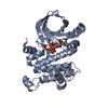
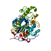
[English] 日本語
Yorodumi
- PDB-3hng: Crystal structure of VEGFR1 in complex with N-(4-Chlorophenyl)-2-... -

+
Open data


ID or keywords:

Loading...

-
Basic information

Entry
Database: PDB / ID: 3hng
TitleCrystal structure of VEGFR1 in complex with N-(4-Chlorophenyl)-2-((pyridin-4-ylmethyl)amino)benzamide
ComponentsVascular endothelial growth factor receptor 1VEGF receptor
KeywordsTRANSFERASE / RECEPTOR TYROSINE KINASE / VEGFR1 / FLT1 / KINASE DOMAIN / INHIBITOR / ACTIVATION LOOP / VASCULAR ENDOTHELIAL GROWTH FACTOR RECEPTOR / STRUCTURAL GENOMICS / STRUCTURAL GENOMICS CONSORTIUM / SGC / SGC STOCKHOLM / Angiogenesis / ATP-binding / Cell membrane / Developmental protein / Differentiation / Disulfide bond / Glycoprotein / Immunoglobulin domain / Kinase / Membrane / Nucleotide-binding / Phosphoprotein / Receptor / Secreted / Transmembrane / Tyrosine-protein kinase
Function / homology
Function and homology information


vascular endothelial growth factor receptor-1 signaling pathway / placental growth factor receptor activity / hyaloid vascular plexus regression / Neurophilin interactions with VEGF and VEGFR / VEGF binds to VEGFR leading to receptor dimerization / vascular endothelial growth factor receptor activity / embryonic morphogenesis / negative regulation of vascular endothelial cell proliferation / blood vessel morphogenesis / growth factor binding ...vascular endothelial growth factor receptor-1 signaling pathway / placental growth factor receptor activity / hyaloid vascular plexus regression / Neurophilin interactions with VEGF and VEGFR / VEGF binds to VEGFR leading to receptor dimerization / vascular endothelial growth factor receptor activity / embryonic morphogenesis / negative regulation of vascular endothelial cell proliferation / blood vessel morphogenesis / growth factor binding / monocyte chemotaxis / cellular response to vascular endothelial growth factor stimulus / vascular endothelial growth factor receptor signaling pathway / positive regulation of phospholipase C activity / transmembrane receptor protein tyrosine kinase activity / positive regulation of MAP kinase activity / receptor protein-tyrosine kinase / peptidyl-tyrosine phosphorylation / cell surface receptor protein tyrosine kinase signaling pathway / positive regulation of angiogenesis / cell migration / actin cytoskeleton / angiogenesis / positive regulation of MAPK cascade / protein autophosphorylation / cell differentiation / positive regulation of phosphatidylinositol 3-kinase/protein kinase B signal transduction / receptor complex / endosome / positive regulation of cell migration / focal adhesion / positive regulation of cell population proliferation / extracellular space / ATP binding / plasma membrane
Similarity search - Function
Vascular endothelial growth factor receptor 1 (VEGFR1) / VEGFR-2, transmembrane domain / VEGFR-2 Transmembrane domain / Vascular endothelial growth factor receptor 1-like, Ig-like domain / Tyrosine-protein kinase, receptor class III, conserved site / Receptor tyrosine kinase class III signature. / Immunoglobulin / Immunoglobulin domain / Immunoglobulin domain / Immunoglobulin I-set ...Vascular endothelial growth factor receptor 1 (VEGFR1) / VEGFR-2, transmembrane domain / VEGFR-2 Transmembrane domain / Vascular endothelial growth factor receptor 1-like, Ig-like domain / Tyrosine-protein kinase, receptor class III, conserved site / Receptor tyrosine kinase class III signature. / Immunoglobulin / Immunoglobulin domain / Immunoglobulin domain / Immunoglobulin I-set / Immunoglobulin I-set domain / Immunoglobulin subtype 2 / Immunoglobulin C-2 Type / Immunoglobulin V-Type / Immunoglobulin V-set domain / Tyrosine-protein kinase, catalytic domain / Tyrosine kinase, catalytic domain / Tyrosine protein kinases specific active-site signature. / Immunoglobulin subtype / Immunoglobulin / Tyrosine-protein kinase, active site / Protein tyrosine and serine/threonine kinase / Serine-threonine/tyrosine-protein kinase, catalytic domain / Transferase(Phosphotransferase) domain 1 / Transferase(Phosphotransferase); domain 1 / Phosphorylase Kinase; domain 1 / Phosphorylase Kinase; domain 1 / Ig-like domain profile. / Immunoglobulin-like domain / Immunoglobulin-like domain superfamily / Protein kinase, ATP binding site / Protein kinases ATP-binding region signature. / Immunoglobulin-like fold / Protein kinase domain profile. / Protein kinase domain / Protein kinase-like domain superfamily / 2-Layer Sandwich / Orthogonal Bundle / Mainly Alpha / Alpha Beta
Similarity search - Domain/homology
Chem-8ST / Vascular endothelial growth factor receptor 1
Similarity search - Component
Biological speciesHomo sapiens (human)
MethodX-RAY DIFFRACTION / SYNCHROTRON / MOLECULAR REPLACEMENT / Resolution: 2.7 Å
AuthorsTresaugues, L. / Roos, A. / Arrowsmith, C.H. / Berglund, H. / Bountra, C. / Collins, R. / Edwards, A.M. / Flodin, S. / Flores, A. / Graslund, S. ...Tresaugues, L. / Roos, A. / Arrowsmith, C.H. / Berglund, H. / Bountra, C. / Collins, R. / Edwards, A.M. / Flodin, S. / Flores, A. / Graslund, S. / Hammarstrom, M. / Johansson, A. / Johansson, I. / Karlberg, T. / Kotenyova, T. / Moche, M. / Nyman, T. / Persson, C. / Kragh-Nielsen, T. / Kotzch, A. / Sagemark, J. / Schueler, H. / Schutz, P. / Siponen, M.I. / Svensson, L. / Thorsell, A.G. / Van der Berg, S. / Weigelt, J. / Welin, M. / Wisniewska, M. / Nordlund, P. / Structural Genomics Consortium (SGC)
CitationJournal: To be Published
Title: Crystal structure of VEGFR1 in complex with N-(4-Chlorophenyl)-2-((pyridin-4-ylmethyl)amino)benzamide
Authors: Tresaugues, L. / Roos, A. / Arrowsmith, C.H. / Berglund, H. / Bountra, C. / Collins, R. / Edwards, A.M. / Flodin, S. / Flores, A. / Graslund, S. / Hammarstrom, M. / Johansson, A. / ...Authors: Tresaugues, L. / Roos, A. / Arrowsmith, C.H. / Berglund, H. / Bountra, C. / Collins, R. / Edwards, A.M. / Flodin, S. / Flores, A. / Graslund, S. / Hammarstrom, M. / Johansson, A. / Johansson, I. / Karlberg, T. / Kotenyova, T. / Moche, M. / Nyman, T. / Persson, C. / Kragh-Nielsen, T. / Kotzch, A. / Sagemark, J. / Schueler, H. / Schutz, P. / Siponen, M.I. / Svensson, L. / Thorsell, A.G. / Van der Berg, S. / Weigelt, J. / Welin, M. / Wisniewska, M. / Nordlund, P.
History
DepositionMay 31, 2009Deposition site: RCSB / Processing site: PDBJ
Revision 1.0Jun 30, 2009Provider: repository / Type: Initial release
Revision 1.1Jul 13, 2011Group: Advisory / Refinement description / Version format compliance
Revision 1.2Nov 1, 2023Group: Data collection / Database references ...Data collection / Database references / Derived calculations / Refinement description
Category: chem_comp_atom / chem_comp_bond ...chem_comp_atom / chem_comp_bond / database_2 / pdbx_initial_refinement_model / struct_ref_seq_dif / struct_site
Item: _database_2.pdbx_DOI / _database_2.pdbx_database_accession ..._database_2.pdbx_DOI / _database_2.pdbx_database_accession / _struct_ref_seq_dif.details / _struct_site.pdbx_auth_asym_id / _struct_site.pdbx_auth_comp_id / _struct_site.pdbx_auth_seq_id

-
Structure visualization

Structure viewerMolecule:
MolmilJmol/JSmol

Downloads & links

-
Assembly

Deposited unit
A: Vascular endothelial growth factor receptor 1
hetero molecules


Theoretical massNumber of molelcules
Total (without water)41,4543
Polymers41,0811
Non-polymers3732
Water59433
1


  • Idetical with deposited unit
  • defined by author&software
TypeNameSymmetry operationNumber
identity operation1_555x,y,z1
MethodPISA
Unit cell
Length a, b, c (Å)58.040, 71.150, 196.020
Angle α, β, γ (deg.)90.00, 90.00, 90.00
Int Tables number23
Space group name H-MI222

-
Components

#1: Protein Vascular endothelial growth factor receptor 1 / VEGF receptor / VEGFR-1 / Vascular permeability factor receptor / Tyrosine-protein kinase receptor FLT / Flt-1 / ...VEGFR-1 / Vascular permeability factor receptor / Tyrosine-protein kinase receptor FLT / Flt-1 / Tyrosine-protein kinase FRT / Fms-like tyrosine kinase 1


Mass: 41081.125 Da / Num. of mol.: 1 / Fragment: KINASE DOMAIN, UNP residues 801-1158
Source method: isolated from a genetically manipulated source
Source: (gene. exp.) Homo sapiens (human) / Gene: FLT1 / Plasmid: pNIC-BSA4 / Production host: Escherichia coli (E. coli) / Strain (production host): BL21(DE3) pRARE
References: UniProt: P17948, receptor protein-tyrosine kinase
#2: Chemical ChemComp-8ST / N-(4-chlorophenyl)-2-[(pyridin-4-ylmethyl)amino]benzamide


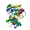
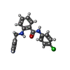
Mass: 337.803 Da / Num. of mol.: 1 / Source method: obtained synthetically / Formula: C19H16ClN3O
#3: Chemical ChemComp-CL / CHLORIDE ION / Chloride


Mass: 35.453 Da / Num. of mol.: 1 / Source method: obtained synthetically / Formula: Cl
#4: Water ChemComp-HOH / water / Water


Mass: 18.015 Da / Num. of mol.: 33 / Source method: isolated from a natural source / Formula: H2O

-
Experimental details

-
Experiment

ExperimentMethod: X-RAY DIFFRACTION / Number of used crystals: 1

-
Sample preparation

CrystalDensity Matthews: 2.46 Å3/Da / Density % sol: 50.06 %
Crystal growTemperature: 277 K / Method: vapor diffusion, sitting drop / pH: 8.5
Details: 0.2M potassium formate, 20% PEG 3350, pH 8.5, VAPOR DIFFUSION, SITTING DROP, temperature 277K

-
Data collection

DiffractionMean temperature: 100 K
Diffraction sourceSource: SYNCHROTRON / Site: ESRF / Beamline: ID14-2 / Wavelength: 0.933 Å
DetectorType: ADSC QUANTUM 4 / Detector: CCD / Date: Apr 4, 2009 / Details: TOROIDAL ZERODUR MIRROR
RadiationMonochromator: DIAMOND (111), GE(220) / Protocol: SINGLE WAVELENGTH / Monochromatic (M) / Laue (L): M / Scattering type: x-ray
Radiation wavelengthWavelength: 0.933 Å / Relative weight: 1
ReflectionResolution: 2.7→44.99 Å / Num. all: 11982 / Num. obs: 11958 / % possible obs: 99.8 % / Redundancy: 4.7 % / Biso Wilson estimate: 65.8 Å2 / Rmerge(I) obs: 0.088 / Rsym value: 0.078 / Net I/σ(I): 13.2
Reflection shellResolution: 2.67→2.81 Å / Redundancy: 4.2 % / Rmerge(I) obs: 0.506 / Mean I/σ(I) obs: 3.2 / Num. unique all: 1669 / Rsym value: 0.441 / % possible all: 98.5

-
Processing

Software
NameVersionClassification
ADSCQuantumdata collection
PHENIXmodel building
REFMAC5.5.0066refinement
iMOSFLMdata reduction
SCALAdata scaling
PHENIXphasing
RefinementMethod to determine structure: MOLECULAR REPLACEMENT
Starting model: PDB ENTRY 2QU5
Resolution: 2.7→44.99 Å / Cor.coef. Fo:Fc: 0.942 / Cor.coef. Fo:Fc free: 0.891 / SU B: 23.981 / SU ML: 0.232 / TLS residual ADP flag: LIKELY RESIDUAL / Isotropic thermal model: ISOTROPIC / Cross valid method: THROUGHOUT / ESU R: 0.654 / ESU R Free: 0.331 / Stereochemistry target values: MAXIMUM LIKELIHOOD / Details: HYDROGENS HAVE BEEN ADDED IN THE RIDING POSITIONS
RfactorNum. reflection% reflectionSelection details
Rfree0.26018 551 4.8 %RANDOM
Rwork0.19431 ---
all0.19739 11021 --
obs0.19739 10998 99.79 %-
Solvent computationIon probe radii: 0.8 Å / Shrinkage radii: 0.8 Å / VDW probe radii: 1.4 Å / Solvent model: MASK
Displacement parametersBiso mean: 26.129 Å2
Baniso -1Baniso -2Baniso -3
1-0.09 Å20 Å20 Å2
2--1.14 Å20 Å2
3----1.23 Å2
Refinement stepCycle: LAST / Resolution: 2.7→44.99 Å
ProteinNucleic acidLigandSolventTotal
Num. atoms2290 0 25 33 2348
Refine LS restraints
Refine-IDTypeDev idealDev ideal targetNumber
X-RAY DIFFRACTIONr_bond_refined_d0.0130.0222374
X-RAY DIFFRACTIONr_bond_other_d0.0010.022186
X-RAY DIFFRACTIONr_angle_refined_deg1.461.9883196
X-RAY DIFFRACTIONr_angle_other_deg0.6435086
X-RAY DIFFRACTIONr_dihedral_angle_1_deg7.0775286
X-RAY DIFFRACTIONr_dihedral_angle_2_deg32.97623.168101
X-RAY DIFFRACTIONr_dihedral_angle_3_deg19.82515433
X-RAY DIFFRACTIONr_dihedral_angle_4_deg20.5081518
X-RAY DIFFRACTIONr_chiral_restr0.0730.2343
X-RAY DIFFRACTIONr_gen_planes_refined0.0070.0212583
X-RAY DIFFRACTIONr_gen_planes_other00.021495
X-RAY DIFFRACTIONr_mcbond_it0.5471.51434
X-RAY DIFFRACTIONr_mcbond_other0.0941.5584
X-RAY DIFFRACTIONr_mcangle_it1.08622304
X-RAY DIFFRACTIONr_scbond_it1.5843940
X-RAY DIFFRACTIONr_scangle_it2.7274.5891
LS refinement shellResolution: 2.7→2.77 Å / Total num. of bins used: 20
RfactorNum. reflection% reflection
Rfree0.37 33 -
Rwork0.335 798 -
obs-798 97.42 %
Refinement TLS params.

Method: refined / Refine-ID: X-RAY DIFFRACTION

IDL112)L122)L132)L222)L232)L332)S11 (Å °)S12 (Å °)S13 (Å °)S21 (Å °)S22 (Å °)S23 (Å °)S31 (Å °)S32 (Å °)S33 (Å °)T112)T122)T132)T222)T232)T332)Origin x (Å)Origin y (Å)Origin z (Å)
15.84345.1424-6.79945.9662-5.80468.29981.5017-0.13790.64331.5686-0.4986-0.7771-2.08990.6879-1.00310.9245-0.3877-0.08291.00440.19771.360212.25533.56134.956
23.1897-0.42020.70731.35920.27634.94520.1324-0.2282-0.00830.0456-0.10850.04120.2883-0.0291-0.02390.0397-0.00470.01040.0860.02770.05823.70416.48637.955
33.95220.28270.67573.7270.33885.74720.00150.57070.052-0.32070.0732-0.19650.22580.7144-0.07470.13640.0289-0.00570.1750.00010.01675.60217.93515.633
Refinement TLS group
IDRefine-IDRefine TLS-IDAuth asym-IDAuth seq-ID
1X-RAY DIFFRACTION1A803 - 818
2X-RAY DIFFRACTION2A819 - 925
3X-RAY DIFFRACTION3A992 - 1158

+
About Yorodumi

-
News

-
Feb 9, 2022. New format data for meta-information of EMDB entries

New format data for meta-information of EMDB entries

  • Version 3 of the EMDB header file is now the official format.
  • The previous official version 1.9 will be removed from the archive.

Related info.:EMDB header

External links:wwPDB to switch to version 3 of the EMDB data model

-
Aug 12, 2020. Covid-19 info

Covid-19 info

URL: https://pdbj.org/emnavi/covid19.php

New page: Covid-19 featured information page in EM Navigator.

Related info.:Covid-19 info / Mar 5, 2020. Novel coronavirus structure data

+
Mar 5, 2020. Novel coronavirus structure data

Novel coronavirus structure data

Related info.:Yorodumi Speices / Aug 12, 2020. Covid-19 info

External links:COVID-19 featured content - PDBj / Molecule of the Month (242):Coronavirus Proteases

+
Jan 31, 2019. EMDB accession codes are about to change! (news from PDBe EMDB page)

EMDB accession codes are about to change! (news from PDBe EMDB page)

  • The allocation of 4 digits for EMDB accession codes will soon come to an end. Whilst these codes will remain in use, new EMDB accession codes will include an additional digit and will expand incrementally as the available range of codes is exhausted. The current 4-digit format prefixed with “EMD-” (i.e. EMD-XXXX) will advance to a 5-digit format (i.e. EMD-XXXXX), and so on. It is currently estimated that the 4-digit codes will be depleted around Spring 2019, at which point the 5-digit format will come into force.
  • The EM Navigator/Yorodumi systems omit the EMD- prefix.

Related info.:Q: What is EMD? / ID/Accession-code notation in Yorodumi/EM Navigator

External links:EMDB Accession Codes are Changing Soon! / Contact to PDBj

+
Jul 12, 2017. Major update of PDB

Major update of PDB

  • wwPDB released updated PDB data conforming to the new PDBx/mmCIF dictionary.
  • This is a major update changing the version number from 4 to 5, and with Remediation, in which all the entries are updated.
  • In this update, many items about electron microscopy experimental information are reorganized (e.g. em_software).
  • Now, EM Navigator and Yorodumi are based on the updated data.

External links:wwPDB Remediation / Enriched Model Files Conforming to OneDep Data Standards Now Available in the PDB FTP Archive

-
Yorodumi

Thousand views of thousand structures

  • Yorodumi is a browser for structure data from EMDB, PDB, SASBDB, etc.
  • This page is also the successor to EM Navigator detail page, and also detail information page/front-end page for Omokage search.
  • The word "yorodu" (or yorozu) is an old Japanese word meaning "ten thousand". "mi" (miru) is to see.

Related info.:EMDB / PDB / SASBDB / Comparison of 3 databanks / Yorodumi Search / Aug 31, 2016. New EM Navigator & Yorodumi / Yorodumi Papers / Jmol/JSmol / Function and homology information / Changes in new EM Navigator and Yorodumi

Read more