Mass: 18.015 Da / Num. of mol.: 37 / Source method: isolated from a natural source / Formula: H2O
-
Experimental details
-
Experiment
Experiment
Method: X-RAY DIFFRACTION / Number of used crystals: 1
-
Sample preparation
Crystal
Density Matthews: 1.98 Å3/Da / Density % sol: 37.83 %
Crystal grow
Temperature: 291 K / Method: vapor diffusion, hanging drop / pH: 7 Details: 20% PEG3350, 0.2M calcium chloride, 0.1M HEPES, pH 7.0, vapor diffusion, hanging drop, temperature 291K
-
Data collection
Diffraction
ID
Mean temperature (K)
Crystal-ID
1
100
1
2
100
1
3
100
1
Diffraction source
Source
Site
Beamline
ID
Wavelength (Å)
SYNCHROTRON
APS
23-ID-B
1
0.91987
SYNCHROTRON
CLSI
08ID-1
2
1.07221
SYNCHROTRON
CLSI
08ID-1
3
1.00645
Detector
Type
ID
Detector
Date
MAR scanner 300 mm plate
1
IMAGE PLATE
Apr 6, 2009
MAR scanner 300 mm plate
2
IMAGE PLATE
May 17, 2009
MAR scanner 300 mm plate
3
IMAGE PLATE
May 16, 2009
Radiation
ID
Protocol
Monochromatic (M) / Laue (L)
Scattering type
Wavelength-ID
1
SINGLEWAVELENGTH
M
x-ray
1
2
SINGLEWAVELENGTH
M
x-ray
1
3
SINGLEWAVELENGTH
M
x-ray
1
Radiation wavelength
ID
Wavelength (Å)
Relative weight
1
0.91987
1
2
1.07221
1
3
1.00645
1
Reflection
Av σ(I) over netI: 44.47 / D res high: 2.8 Å / D res low: 30 Å
Redundancy (%)
ID
Number
Rmerge(I) obs
Χ2
Num. obs
% possible obs
7.6
1
300995
0.058
1.66
39689
100
12.1
2
259196
0.061
1.76
21389
98.8
Diffraction reflection shell
Highest resolution (Å)
Lowest resolution (Å)
% possible obs (%)
ID
Rmerge(I) obs
Chi squared
Redundancy
6.02
30
99.9
1
0.039
2.188
7.8
4.78
6.02
100
1
0.051
2.515
7.7
4.18
4.78
100
1
0.05
2.115
7.8
3.8
4.18
100
1
0.055
1.787
7.8
3.53
3.8
100
1
0.067
1.812
7.8
3.32
3.53
100
1
0.084
1.406
7.8
3.15
3.32
100
1
0.112
1.251
7.8
3.02
3.15
100
1
0.186
1.132
7.7
2.9
3.02
100
1
0.282
1.149
7.3
2.8
2.9
99.6
1
0.365
1.086
6.2
6.02
30
99.9
2
0.035
2.025
13.2
4.78
6.02
100
2
0.046
2.044
13.9
4.18
4.78
100
2
0.052
2.032
14.1
3.8
4.18
100
2
0.071
1.829
14
3.53
3.8
100
2
0.104
2.058
13.7
3.32
3.53
100
2
0.15
1.686
13.5
3.15
3.32
100
2
0.21
1.461
13.3
3.02
3.15
100
2
0.364
1.249
12.4
2.9
3.02
99.5
2
0.499
1.21
8
2.8
2.9
88.3
2
0.516
1.064
3.6
Reflection
Resolution: 1.8→40 Å / Num. obs: 9198 / % possible obs: 96.8 % / Redundancy: 17 % / Rmerge(I) obs: 0.079 / Χ2: 1.434 / Net I/σ(I): 44.467
Reflection shell
Resolution (Å)
Redundancy (%)
Rmerge(I) obs
Num. unique all
Χ2
% possible all
1.8-1.86
6.2
0.47
693
0.772
75.6
1.86-1.94
10.6
0.364
833
0.795
92.8
1.94-2.03
15.5
0.318
912
0.849
99.2
2.03-2.13
19.2
0.233
897
0.969
99.9
2.13-2.27
20
0.168
933
1.139
100
2.27-2.44
20.1
0.141
930
1.4
100
2.44-2.69
19.9
0.115
932
1.387
100
2.69-3.08
19.6
0.08
960
1.602
100
3.08-3.88
18.7
0.063
980
2.501
99.8
3.88-40
16.7
0.042
1128
1.884
99.3
-
Phasing
Phasing
Method: SIRAS
-
Processing
Software
Name
Version
Classification
NB
DENZO
datareduction
SCALEPACK
datascaling
SHELX
phasing
RESOLVE
phasing
REFMAC
5.5.0072
refinement
PDB_EXTRACT
3.006
dataextraction
HKL-2000
datareduction
HKL-2000
datascaling
Refinement
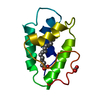
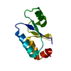
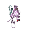
Method to determine structure: SIRAS / Resolution: 1.8→19.905 Å / Cor.coef. Fo:Fc: 0.937 / Cor.coef. Fo:Fc free: 0.898 / WRfactor Rfree: 0.254 / WRfactor Rwork: 0.213 / Occupancy max: 1 / Occupancy min: 0.01 / SU B: 2.851 / SU ML: 0.09 / Cross valid method: THROUGHOUT / σ(F): 0 / ESU R: 0.141 / ESU R Free: 0.143 / Stereochemistry target values: MAXIMUM LIKELIHOOD Details: HYDROGENS HAVE BEEN ADDED IN THE RIDING POSITIONS U VALUES : REFINED INDIVIDUALLY. Structure was solved in I4122 spacegroup using SIRAS, using a thimerosal-soaked sample as "native" and a ...Details: HYDROGENS HAVE BEEN ADDED IN THE RIDING POSITIONS U VALUES : REFINED INDIVIDUALLY. Structure was solved in I4122 spacegroup using SIRAS, using a thimerosal-soaked sample as "native" and a tetrabromoplatinate derivative. DM and RESOLVE were used for NCS averaging after a rough outline of the protein chain was traced in the initial map. Preliminary model was placed into refinement space group using PHASER. The model was automatically updated with ARP/WARP and further refined/validated wit REFMAC/MOLPROBITY.
Rfactor
Num. reflection
% reflection
Selection details
Rfree
0.275
433
4.769 %
RANDOM
Rwork
0.223
-
-
-
obs
0.225
9080
97.04 %
-
Solvent computation
Ion probe radii: 0.8 Å / Shrinkage radii: 0.8 Å / VDW probe radii: 1.4 Å / Solvent model: MASK BULK SOLVENT
In the structure databanks used in Yorodumi, some data are registered as the other names, "COVID-19 virus" and "2019-nCoV". Here are the details of the virus and the list of structure data.
Jan 31, 2019. EMDB accession codes are about to change! (news from PDBe EMDB page)
EMDB accession codes are about to change! (news from PDBe EMDB page)
The allocation of 4 digits for EMDB accession codes will soon come to an end. Whilst these codes will remain in use, new EMDB accession codes will include an additional digit and will expand incrementally as the available range of codes is exhausted. The current 4-digit format prefixed with “EMD-” (i.e. EMD-XXXX) will advance to a 5-digit format (i.e. EMD-XXXXX), and so on. It is currently estimated that the 4-digit codes will be depleted around Spring 2019, at which point the 5-digit format will come into force.
The EM Navigator/Yorodumi systems omit the EMD- prefix.
Related info.:Q: What is EMD? / ID/Accession-code notation in Yorodumi/EM Navigator
Yorodumi is a browser for structure data from EMDB, PDB, SASBDB, etc.
This page is also the successor to EM Navigator detail page, and also detail information page/front-end page for Omokage search.
The word "yorodu" (or yorozu) is an old Japanese word meaning "ten thousand". "mi" (miru) is to see.
Related info.:EMDB / PDB / SASBDB / Comparison of 3 databanks / Yorodumi Search / Aug 31, 2016. New EM Navigator & Yorodumi / Yorodumi Papers / Jmol/JSmol / Function and homology information / Changes in new EM Navigator and Yorodumi
 Movie
Movie Controller

 Yorodumi
 Open data
 Basic information
 Components
 Keywords
 Function and homology information
 Homo sapiens (human)
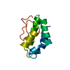
 X-RAY DIFFRACTION /
  Authors
 Citation
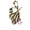
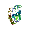
 Structure visualization
 Downloads & links
 Other downloads
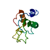

 PDBj
PDBj
 Assembly


 Mass: 40.078 Da / Num. of mol.: 1 / Source method: obtained synthetically / Formula: Ca
 Num. of mol.: 5 / Source method: obtained synthetically
 Mass: 18.015 Da / Num. of mol.: 37 / Source method: isolated from a natural source / Formula: H2O
 Sample preparation

 Processing