 Movie
Movie Controller

[English] 日本語
 Yorodumi
- PDB-3hef: Crystal structure of the bacteriophage Sf6 terminase small subunit -
+ Open data

- Basic information
| Entry | Database: PDB / ID: 3hef | ||||||
|---|---|---|---|---|---|---|---|
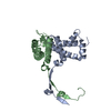
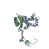
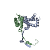
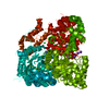
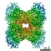
| Title | Crystal structure of the bacteriophage Sf6 terminase small subunit | ||||||
 Components Components | Gene 1 protein | ||||||
 Keywords Keywords | VIRAL PROTEIN / bacteriophage Sf6 / terminase small subunit gp1 / gp1 octameric assembly / gp1 channel / DNA recognition / DNA packaging | ||||||
| Function / homology | : / Bacteriophage Sf6, terminase small subunit-like / Homeodomain-like / Arc Repressor Mutant, subunit A / Orthogonal Bundle / Mainly Alpha / identical protein binding / Gene 1 protein Function and homology information Function and homology information | ||||||
| Biological species |  Enterobacteria phage Sf6 (virus) Enterobacteria phage Sf6 (virus) | ||||||
| Method |  X-RAY DIFFRACTION / SYNCHROTRON / MAD / Resolution: 1.65 Å X-RAY DIFFRACTION / SYNCHROTRON / MAD / Resolution: 1.65 Å | ||||||
 Authors Authors | Zhao, H. / Tang, L. | ||||||
 Citation Citation | Journal: Proc.Natl.Acad.Sci.USA / Year: 2010 Title: Crystal structure of the DNA-recognition component of the bacterial virus Sf6 genome-packaging machine Authors: Zhao, H. / Finch, C.J. / Sequeira, R.D. / Johnson, B.A. / Johnson, J.E. / Casjens, S.R. / Tang, L. | ||||||
| History |
|
- Structure visualization
| Structure viewer | Molecule: MolmilJmol/JSmol |
|---|
- Downloads & links
-Download
| PDBx/mmCIF format | 3hef.cif.gz | 67.2 KB | Display | PDBx/mmCIF format |
|---|---|---|---|---|
| PDB format | pdb3hef.ent.gz | 49.1 KB | Display | PDB format |
| PDBx/mmJSON format | 3hef.json.gz | Tree view | PDBx/mmJSON format | |
| Others |  Other downloads Other downloads |
-Validation report
| Arichive directory | https://data.pdbj.org/pub/pdb/validation_reports/he/3hefftp://data.pdbj.org/pub/pdb/validation_reports/he/3hef | HTTPS FTP |
|---|
-Related structure data
| Similar structure data |
|---|


-Links
 PDBj
PDBj- Assembly
| Deposited unit | 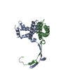
| ||||||||
|---|---|---|---|---|---|---|---|---|---|
| 1 | 
| ||||||||
| Unit cell |
|
-Components
| #1: Protein | Mass: 15859.057 Da / Num. of mol.: 2 Source method: isolated from a genetically manipulated source Source: (gene. exp.) Enterobacteria phage Sf6 (virus) / Gene: gp1 / Plasmid: pET28b / Production host:  #2: Water | ChemComp-HOH / |  Mass: 18.015 Da / Num. of mol.: 254 / Source method: isolated from a natural source / Formula: H2O Mass: 18.015 Da / Num. of mol.: 254 / Source method: isolated from a natural source / Formula: H2O |
|---|
-Experimental details
-Experiment
| Experiment | Method: X-RAY DIFFRACTION / Number of used crystals: 1 |
|---|
- Sample preparation
| Crystal | Density Matthews: 2.26 Å3/Da / Density % sol: 45.58 % |
|---|---|
| Crystal grow | Temperature: 293 K / Method: vapor diffusion, hanging drop / pH: 7 Details: 1.2M NaH2PO4/0.8M K2HPO4, 0.2M Li2SO4, 5% glycerol, pH 7.0, VAPOR DIFFUSION, HANGING DROP, temperature 293K |
-Data collection
| Diffraction |
| |||||||||||||||||||||||||||||||||||||||||||||||||||||||||||||||||||||||||||||
|---|---|---|---|---|---|---|---|---|---|---|---|---|---|---|---|---|---|---|---|---|---|---|---|---|---|---|---|---|---|---|---|---|---|---|---|---|---|---|---|---|---|---|---|---|---|---|---|---|---|---|---|---|---|---|---|---|---|---|---|---|---|---|---|---|---|---|---|---|---|---|---|---|---|---|---|---|---|---|
| Diffraction source |
| |||||||||||||||||||||||||||||||||||||||||||||||||||||||||||||||||||||||||||||
| Detector |
| |||||||||||||||||||||||||||||||||||||||||||||||||||||||||||||||||||||||||||||
| Radiation |
| |||||||||||||||||||||||||||||||||||||||||||||||||||||||||||||||||||||||||||||
| Radiation wavelength |
| |||||||||||||||||||||||||||||||||||||||||||||||||||||||||||||||||||||||||||||
| Reflection | Redundancy: 7.2 % / Av σ(I) over netI: 107.12 / Number: 176519 / Rmerge(I) obs: 0.065 / Χ2: 1.8 / D res high: 1.86 Å / D res low: 50 Å / Num. obs: 24582 / % possible obs: 98.7 | |||||||||||||||||||||||||||||||||||||||||||||||||||||||||||||||||||||||||||||
| Diffraction reflection shell |
| |||||||||||||||||||||||||||||||||||||||||||||||||||||||||||||||||||||||||||||
| Reflection | Resolution: 1.65→50 Å / Num. all: 35754 / Num. obs: 35647 / % possible obs: 99.7 % / Observed criterion σ(F): 2 / Observed criterion σ(I): 2 / Redundancy: 28.4 % / Rmerge(I) obs: 0.053 / Χ2: 1.925 / Net I/σ(I): 107.121 | |||||||||||||||||||||||||||||||||||||||||||||||||||||||||||||||||||||||||||||
| Reflection shell | Resolution: 1.65→1.71 Å / Redundancy: 28.6 % / Rmerge(I) obs: 0.521 / Num. unique all: 3521 / Χ2: 1.223 / % possible all: 100 |

-Phasing
| Phasing dm | FOM : 0.57 / FOM acentric: 0.56 / FOM centric: 0.61 / Reflection: 24251 / Reflection acentric: 21281 / Reflection centric: 2970 | |||||||||||||||||||||||||||||||||||||||||||||||||
|---|---|---|---|---|---|---|---|---|---|---|---|---|---|---|---|---|---|---|---|---|---|---|---|---|---|---|---|---|---|---|---|---|---|---|---|---|---|---|---|---|---|---|---|---|---|---|---|---|---|---|
| Phasing dm shell |
|
- Processing
| Software |
| ||||||||||||||||||||||||||||||||
|---|---|---|---|---|---|---|---|---|---|---|---|---|---|---|---|---|---|---|---|---|---|---|---|---|---|---|---|---|---|---|---|---|---|
| Refinement | Method to determine structure: MAD / Resolution: 1.65→20 Å / Occupancy max: 1 / Occupancy min: 1 / FOM work R set: 0.851 / σ(F): 1037 / Stereochemistry target values: Engh & Huber
| ||||||||||||||||||||||||||||||||
| Solvent computation | Bsol: 63.494 Å2 | ||||||||||||||||||||||||||||||||
| Displacement parameters | Biso max: 95.21 Å2 / Biso mean: 35.237 Å2 / Biso min: 11.69 Å2
| ||||||||||||||||||||||||||||||||
| Refine analyze |
| ||||||||||||||||||||||||||||||||
| Refinement step | Cycle: LAST / Resolution: 1.65→20 Å
| ||||||||||||||||||||||||||||||||
| Refine LS restraints |
| ||||||||||||||||||||||||||||||||
| LS refinement shell | Resolution: 1.65→1.75 Å / Rfactor Rfree error: 0.019
| ||||||||||||||||||||||||||||||||
| Xplor file |
|
