 Movie
Movie Controller

[English] 日本語
 Yorodumi
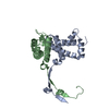
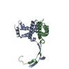
- PDB-4dzp: Crystal structure of the terminase small subunit gp1 with R48A mu... -
+ Open data

- Basic information
| Entry | Database: PDB / ID: 4dzp | ||||||
|---|---|---|---|---|---|---|---|
| Title | Crystal structure of the terminase small subunit gp1 with R48A mutation of the bacterial virus sf6 | ||||||
 Components Components | Gene 1 protein | ||||||
 Keywords Keywords | VIRAL PROTEIN / gp1 / DNA-binding / R48A mutation | ||||||
| Function / homology | : / Bacteriophage Sf6, terminase small subunit-like / Homeodomain-like / Arc Repressor Mutant, subunit A / Orthogonal Bundle / Mainly Alpha / identical protein binding / Gene 1 protein Function and homology information Function and homology information | ||||||
| Biological species |  Shigella phage Sf6 (virus) Shigella phage Sf6 (virus) | ||||||
| Method |  X-RAY DIFFRACTION / SYNCHROTRON / MOLECULAR REPLACEMENT / Resolution: 1.8 Å X-RAY DIFFRACTION / SYNCHROTRON / MOLECULAR REPLACEMENT / Resolution: 1.8 Å | ||||||
 Authors Authors | Zhao, H. / Tang, L. | ||||||
 Citation Citation | Journal: J.Mol.Biol. / Year: 2012 Title: Structural and Functional Studies of the Phage Sf6 Terminase Small Subunit Reveal a DNA-Spooling Device Facilitated by Structural Plasticity. Authors: Zhao, H. / Kamau, Y.N. / Christensen, T.E. / Tang, L. | ||||||
| History |
|
- Structure visualization
| Structure viewer | Molecule: MolmilJmol/JSmol |
|---|
- Downloads & links
-Download
| PDBx/mmCIF format | 4dzp.cif.gz | 65.3 KB | Display | PDBx/mmCIF format |
|---|---|---|---|---|
| PDB format | pdb4dzp.ent.gz | 48.4 KB | Display | PDB format |
| PDBx/mmJSON format | 4dzp.json.gz | Tree view | PDBx/mmJSON format | |
| Others |  Other downloads Other downloads |
-Validation report
| Arichive directory | https://data.pdbj.org/pub/pdb/validation_reports/dz/4dzpftp://data.pdbj.org/pub/pdb/validation_reports/dz/4dzp | HTTPS FTP |
|---|
-Related structure data
| Related structure data |  4dycC 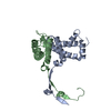 4dyqC  4dyrC 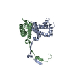 4dzjC  3hefS S: Starting model for refinement C: citing same article ( |
|---|---|
| Similar structure data |


-Links
 PDBj
PDBj- Assembly
| Deposited unit | 
| ||||||||
|---|---|---|---|---|---|---|---|---|---|
| 1 | 
| ||||||||
| Unit cell |
| ||||||||
| Components on special symmetry positions |
|
-Components
| #1: Protein | Mass: 15490.663 Da / Num. of mol.: 2 / Mutation: R48A Source method: isolated from a genetically manipulated source Source: (gene. exp.) Shigella phage Sf6 (virus) / Strain: bacteriophage Sf6 / Gene: gp1 / Plasmid: pET20b / Production host:  #2: Water | ChemComp-HOH / |  Mass: 18.015 Da / Num. of mol.: 213 / Source method: isolated from a natural source / Formula: H2O Mass: 18.015 Da / Num. of mol.: 213 / Source method: isolated from a natural source / Formula: H2O |
|---|
-Experimental details
-Experiment
| Experiment | Method: X-RAY DIFFRACTION / Number of used crystals: 1 |
|---|
- Sample preparation
| Crystal | Density Matthews: 2.33 Å3/Da / Density % sol: 47.2 % |
|---|---|
| Crystal grow | Temperature: 293 K / Method: vapor diffusion, hanging drop / pH: 6.4 Details: 1.2M NAH2PO4/0.8M K2HPO4, 0.2M Li2SO4, pH 6.4, VAPOR DIFFUSION, HANGING DROP, temperature 293K |
-Data collection
| Diffraction | Mean temperature: 298 K |
|---|---|
| Diffraction source | Source: SYNCHROTRON / Site: SSRL  / Beamline: BL12-2 / Wavelength: 0.9795 Å / Beamline: BL12-2 / Wavelength: 0.9795 Å |
| Detector | Type: PSI PILATUS 6M / Detector: PIXEL / Date: Mar 27, 2010 / Details: Rh coated |
| Radiation | Monochromator: Liquid nitrogen-cooled double crystal / Protocol: SINGLE WAVELENGTH / Monochromatic (M) / Laue (L): M / Scattering type: x-ray |
| Radiation wavelength | Wavelength: 0.9795 Å / Relative weight: 1 |
| Reflection | Resolution: 1.8→19.221 Å / Num. all: 27730 / Num. obs: 27623 / % possible obs: 99.6 % / Observed criterion σ(F): 0 / Observed criterion σ(I): -3 / Redundancy: 8.38 % / Biso Wilson estimate: 26.637 Å2 / Rmerge(I) obs: 0.034 / Net I/σ(I): 31.71 |
| Reflection shell | Resolution: 1.8→1.85 Å / Redundancy: 8.78 % / Rmerge(I) obs: 0.203 / Mean I/σ(I) obs: 7.78 / Num. unique all: 1977 / % possible all: 98.9 |
- Processing
| Software |
| |||||||||||||||||||||||||||||||||||||||||||||||||||||||||||||||||||||||||||||
|---|---|---|---|---|---|---|---|---|---|---|---|---|---|---|---|---|---|---|---|---|---|---|---|---|---|---|---|---|---|---|---|---|---|---|---|---|---|---|---|---|---|---|---|---|---|---|---|---|---|---|---|---|---|---|---|---|---|---|---|---|---|---|---|---|---|---|---|---|---|---|---|---|---|---|---|---|---|---|
| Refinement | Method to determine structure: MOLECULAR REPLACEMENT Starting model: PDB code 3HEF Resolution: 1.8→19.221 Å / SU ML: 0.01 / Isotropic thermal model: anisotropic / Cross valid method: THROUGHOUT / σ(F): 1.36 / Phase error: 22.72 / Stereochemistry target values: ML
| |||||||||||||||||||||||||||||||||||||||||||||||||||||||||||||||||||||||||||||
| Solvent computation | Shrinkage radii: 0.9 Å / VDW probe radii: 1.11 Å / Solvent model: FLAT BULK SOLVENT MODEL / Bsol: 46.394 Å2 / ksol: 0.369 e/Å3 | |||||||||||||||||||||||||||||||||||||||||||||||||||||||||||||||||||||||||||||
| Displacement parameters | Biso mean: 35.88 Å2
| |||||||||||||||||||||||||||||||||||||||||||||||||||||||||||||||||||||||||||||
| Refinement step | Cycle: LAST / Resolution: 1.8→19.221 Å
| |||||||||||||||||||||||||||||||||||||||||||||||||||||||||||||||||||||||||||||
| Refine LS restraints |
| |||||||||||||||||||||||||||||||||||||||||||||||||||||||||||||||||||||||||||||
| LS refinement shell |
|
