 Movie
Movie Controller

[English] 日本語
 Yorodumi
- PDB-3dbl: Structural Dissection of a Gating Mechanism Preventing Misactivat... -
+ Open data

- Basic information
| Entry | Database: PDB / ID: 3dbl | ||||||
|---|---|---|---|---|---|---|---|
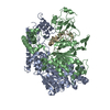
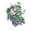
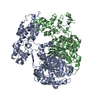
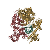
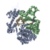
| Title | Structural Dissection of a Gating Mechanism Preventing Misactivation of Ubiquitin by NEDD8's E1 (APPBP1-UBA3Arg190wt-NEDD8Ala72Gln) | ||||||
 Components Components |
| ||||||
 Keywords Keywords | CELL CYCLE / activating enzyme / Apoptosis / Membrane / Ubl conjugation pathway / ATP-binding / Ligase / Nucleotide-binding / Polymorphism / Nucleus | ||||||
| Function / homology |  Function and homology information Function and homology informationE1 NEDD8-activating enzyme / NEDD8 activating enzyme activity / endomitotic cell cycle / cellular response to camptothecin / NEDD8 transferase activity / mitotic DNA replication checkpoint signaling / protein neddylation / TGF-beta receptor signaling activates SMADs / regulation of proteolysis / regulation of postsynapse assembly ...E1 NEDD8-activating enzyme / NEDD8 activating enzyme activity / endomitotic cell cycle / cellular response to camptothecin / NEDD8 transferase activity / mitotic DNA replication checkpoint signaling / protein neddylation / TGF-beta receptor signaling activates SMADs / regulation of proteolysis / regulation of postsynapse assembly / anatomical structure morphogenesis / post-translational protein modification / regulation of neuron apoptotic process / protein modification process / NIK-->noncanonical NF-kB signaling / Dectin-1 mediated noncanonical NF-kB signaling / Iron uptake and transport / modification-dependent protein catabolic process / protein tag activity / intracellular protein localization / UCH proteinases / Cargo recognition for clathrin-mediated endocytosis / Antigen processing: Ubiquitination & Proteasome degradation / Neddylation / neuron apoptotic process / regulation of apoptotic process / ubiquitin-dependent protein catabolic process / regulation of cell cycle / postsynapse / protein heterodimerization activity / ubiquitin protein ligase binding / regulation of transcription by RNA polymerase II / glutamatergic synapse / signal transduction / protein-containing complex / proteolysis / extracellular exosome / nucleoplasm / ATP binding / identical protein binding / nucleus / plasma membrane / cytoplasm / cytosol Similarity search - Function | ||||||
| Biological species |  Homo sapiens (human) Homo sapiens (human) | ||||||
| Method |  X-RAY DIFFRACTION / SYNCHROTRON / rigid body refinement / Resolution: 2.9 Å X-RAY DIFFRACTION / SYNCHROTRON / rigid body refinement / Resolution: 2.9 Å | ||||||
 Authors Authors | Souphron, J. / Schulman, B.A. | ||||||
 Citation Citation | Journal: Biochemistry / Year: 2008 Title: Structural dissection of a gating mechanism preventing misactivation of ubiquitin by NEDD8's E1. Authors: Souphron, J. / Waddell, M.B. / Paydar, A. / Tokgoz-Gromley, Z. / Roussel, M.F. / Schulman, B.A. | ||||||
| History |
|
- Structure visualization
| Structure viewer | Molecule: MolmilJmol/JSmol |
|---|
- Downloads & links
-Download
| PDBx/mmCIF format | 3dbl.cif.gz | 798.6 KB | Display | PDBx/mmCIF format |
|---|---|---|---|---|
| PDB format | pdb3dbl.ent.gz | 650.1 KB | Display | PDB format |
| PDBx/mmJSON format | 3dbl.json.gz | Tree view | PDBx/mmJSON format | |
| Others |  Other downloads Other downloads |
-Validation report
| Arichive directory | https://data.pdbj.org/pub/pdb/validation_reports/db/3dblftp://data.pdbj.org/pub/pdb/validation_reports/db/3dbl | HTTPS FTP |
|---|
-Related structure data
| Related structure data | 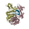 3dbhSC 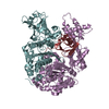 3dbrC S: Starting model for refinement C: citing same article ( |
|---|---|
| Similar structure data |


-Links
 PDBj
PDBj


- Assembly
| Deposited unit | 
| ||||||||
|---|---|---|---|---|---|---|---|---|---|
| 1 | 
| ||||||||
| 2 | 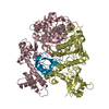
| ||||||||
| 3 | 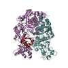
| ||||||||
| 4 | 
| ||||||||
| Unit cell |
|
-Components
| #1: Protein | Mass: 59973.781 Da / Num. of mol.: 4 Source method: isolated from a genetically manipulated source Source: (gene. exp.) Homo sapiens (human) / Gene: NAE1, APPBP1 / Plasmid: pABLO / Production host:  #2: Protein | Mass: 48767.750 Da / Num. of mol.: 4 / Mutation: C216A Source method: isolated from a genetically manipulated source Source: (gene. exp.) Homo sapiens (human) / Gene: UBA3, UBE1C / Plasmid: pABLO / Production host: References: UniProt: Q8TBC4, Ligases; Forming carbon-nitrogen bonds; Acid-amino-acid ligases (peptide synthases) #3: Protein | Mass: 9692.143 Da / Num. of mol.: 4 / Mutation: A172Q Source method: isolated from a genetically manipulated source Source: (gene. exp.) Homo sapiens (human) / Gene: NEDD8 / Plasmid: pGEX2TK / Production host: #4: Chemical | ChemComp-ZN /   Mass: 65.409 Da / Num. of mol.: 4 / Source method: obtained synthetically / Formula: Zn Mass: 65.409 Da / Num. of mol.: 4 / Source method: obtained synthetically / Formula: Zn |
|---|
-Experimental details
-Experiment
| Experiment | Method: X-RAY DIFFRACTION / Number of used crystals: 1 |
|---|
- Sample preparation
| Crystal | Density Matthews: 3 Å3/Da / Density % sol: 59.02 % |
|---|---|
| Crystal grow | Temperature: 291 K / Method: vapor diffusion, hanging drop / pH: 7.5 Details: 0.1 M Tris, 0.4 M ammonium acetate, 9-10% PEG 10K, 5 mM DTT, pH 7.5, VAPOR DIFFUSION, HANGING DROP, temperature 291K |
-Data collection
| Diffraction source | Source: SYNCHROTRON / Site: ALS  / Beamline: 8.2.2 / Wavelength: 1 Å / Beamline: 8.2.2 / Wavelength: 1 Å |
|---|---|
| Detector | Type: ADSC QUANTUM 315 / Detector: CCD / Date: Aug 26, 2006 / Details: mirrors |
| Radiation | Protocol: SINGLE WAVELENGTH / Monochromatic (M) / Laue (L): M / Scattering type: x-ray |
| Radiation wavelength | Wavelength: 1 Å / Relative weight: 1 |
| Reflection | Resolution: 2.9→50 Å / Num. all: 117851 / Num. obs: 116696 / % possible obs: 93.7 % / Redundancy: 4.9 % / Rmerge(I) obs: 0.163 / Net I/σ(I): 13.2 |
| Reflection shell | Resolution: 2.9→3 Å / Redundancy: 2.6 % / Rmerge(I) obs: 0.468 / Mean I/σ(I) obs: 1.8 / Num. unique all: 9776 / % possible all: 78.6 |
- Processing
| Software |
| |||||||||||||||||||||||||
|---|---|---|---|---|---|---|---|---|---|---|---|---|---|---|---|---|---|---|---|---|---|---|---|---|---|---|
| Refinement | Method to determine structure: rigid body refinement Starting model: PDB entry 3DBH Resolution: 2.9→50 Å / Cross valid method: THROUGHOUT / Stereochemistry target values: Engh & Huber
| |||||||||||||||||||||||||
| Displacement parameters | Biso mean: 22.26 Å2
| |||||||||||||||||||||||||
| Refinement step | Cycle: LAST / Resolution: 2.9→50 Å
| |||||||||||||||||||||||||
| Refine LS restraints |
|
