 Movie
Movie Controller

[English] 日本語
 Yorodumi
- PDB-3he7: Crystal structure of mouse CD1d-alpha-galactosylceramide with mou... -
+ Open data

- Basic information
| Entry | Database: PDB / ID: 3he7 | |||||||||
|---|---|---|---|---|---|---|---|---|---|---|
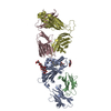
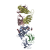
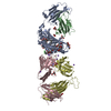
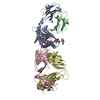
| Title | Crystal structure of mouse CD1d-alpha-galactosylceramide with mouse Valpha14-Vbeta7 NKT TCR | |||||||||
 Components Components |
| |||||||||
 Keywords Keywords | IMMUNE SYSTEM / mouse CD1d / mouse NKT T-cell receptors / Cell membrane / Disulfide bond / Endosome / Glycoprotein / Immune response / Immunoglobulin domain / Innate immunity / Lysosome / Membrane / Transmembrane / MHC I | |||||||||
| Function / homology |  Function and homology information Function and homology informationregulation of immature T cell proliferation in thymus / lipid antigen binding / positive regulation of NK T cell activation / positive regulation of NK T cell differentiation / NK T cell differentiation / endogenous lipid antigen binding / exogenous lipid antigen binding / antigen processing and presentation, endogenous lipid antigen via MHC class Ib / lipopeptide binding / antigen processing and presentation, exogenous lipid antigen via MHC class Ib ...regulation of immature T cell proliferation in thymus / lipid antigen binding / positive regulation of NK T cell activation / positive regulation of NK T cell differentiation / NK T cell differentiation / endogenous lipid antigen binding / exogenous lipid antigen binding / antigen processing and presentation, endogenous lipid antigen via MHC class Ib / lipopeptide binding / antigen processing and presentation, exogenous lipid antigen via MHC class Ib / positive thymic T cell selection / positive regulation of macrophage activation / Endosomal/Vacuolar pathway / DAP12 interactions / Antigen Presentation: Folding, assembly and peptide loading of class I MHC / ER-Phagosome pathway / DAP12 signaling / alpha-beta T cell receptor complex / Immunoregulatory interactions between a Lymphoid and a non-Lymphoid cell / antigen processing and presentation / Translocation of ZAP-70 to Immunological synapse / Phosphorylation of CD3 and TCR zeta chains / regulation of membrane depolarization / positive regulation of interleukin-4 production / alpha-beta T cell activation / Generation of second messenger molecules / Co-inhibition by PD-1 / regulation of immune response / positive regulation of interleukin-2 production / cellular defense response / T cell receptor binding / Neutrophil degranulation / positive regulation of T cell proliferation / cell adhesion molecule binding / response to bacterium / regulation of iron ion transport / cellular response to iron(III) ion / negative regulation of iron ion transport / negative regulation of forebrain neuron differentiation / antigen processing and presentation of exogenous protein antigen via MHC class Ib, TAP-dependent / iron ion transport / peptide antigen assembly with MHC class I protein complex / regulation of erythrocyte differentiation / response to molecule of bacterial origin / HFE-transferrin receptor complex / MHC class I peptide loading complex / transferrin transport / cellular response to iron ion / negative regulation of receptor-mediated endocytosis / positive regulation of T cell cytokine production / antigen processing and presentation of endogenous peptide antigen via MHC class I / MHC class I protein complex / peptide antigen assembly with MHC class II protein complex / negative regulation of neurogenesis / MHC class II protein complex / cellular response to nicotine / positive regulation of receptor-mediated endocytosis / multicellular organismal-level iron ion homeostasis / positive regulation of T cell mediated cytotoxicity / antigen processing and presentation of exogenous peptide antigen via MHC class II / positive regulation of immune response / peptide antigen binding / positive regulation of type II interferon production / phagocytic vesicle membrane / positive regulation of T cell activation / negative regulation of epithelial cell proliferation / Immunoregulatory interactions between a Lymphoid and a non-Lymphoid cell / sensory perception of smell / positive regulation of cellular senescence / MHC class II protein complex binding / T cell differentiation in thymus / late endosome / late endosome membrane / Downstream TCR signaling / antimicrobial humoral immune response mediated by antimicrobial peptide / negative regulation of neuron projection development / T cell receptor signaling pathway / antibacterial humoral response / protein refolding / cellular response to lipopolysaccharide / amyloid fibril formation / protein homotetramerization / defense response to Gram-negative bacterium / intracellular iron ion homeostasis / adaptive immune response / learning or memory / early endosome / lysosome / endosome membrane / defense response to Gram-positive bacterium / immune response / external side of plasma membrane / innate immune response / lysosomal membrane / structural molecule activity / cell surface / Golgi apparatus / endoplasmic reticulum / protein homodimerization activity / : Similarity search - Function | |||||||||
| Biological species |  | |||||||||
| Method |  X-RAY DIFFRACTION / SYNCHROTRON / MOLECULAR REPLACEMENT / Resolution: 2.8 Å X-RAY DIFFRACTION / SYNCHROTRON / MOLECULAR REPLACEMENT / Resolution: 2.8 Å | |||||||||
 Authors Authors | Patel, O. / Rossjohn, J. | |||||||||
 Citation Citation | Journal: Immunity / Year: 2009 Title: Differential recognition of CD1d-alpha-galactosyl ceramide by the V beta 8.2 and V beta 7 semi-invariant NKT T cell receptors Authors: Pellicci, D.G. / Patel, O. / Kjer-Nielsen, L. / Pang, S.S. / Sullivan, L.C. / Kyparissoudis, K. / Brooks, A.G. / Reid, H.H. / Gras, S. / Lucet, I.S. / Koh, R. / Smyth, M.J. / Mallevaey, T. / ...Authors: Pellicci, D.G. / Patel, O. / Kjer-Nielsen, L. / Pang, S.S. / Sullivan, L.C. / Kyparissoudis, K. / Brooks, A.G. / Reid, H.H. / Gras, S. / Lucet, I.S. / Koh, R. / Smyth, M.J. / Mallevaey, T. / Matsuda, J.L. / Gapin, L. / McCluskey, J. / Godfrey, D.I. / Rossjohn, J. | |||||||||
| History |
|
- Structure visualization
| Structure viewer | Molecule: MolmilJmol/JSmol |
|---|
- Downloads & links
-Download
| PDBx/mmCIF format | 3he7.cif.gz | 178.7 KB | Display | PDBx/mmCIF format |
|---|---|---|---|---|
| PDB format | pdb3he7.ent.gz | 137.9 KB | Display | PDB format |
| PDBx/mmJSON format | 3he7.json.gz | Tree view | PDBx/mmJSON format | |
| Others |  Other downloads Other downloads |
-Validation report
| Arichive directory | https://data.pdbj.org/pub/pdb/validation_reports/he/3he7ftp://data.pdbj.org/pub/pdb/validation_reports/he/3he7 | HTTPS FTP |
|---|
-Related structure data
| Related structure data |  3he6SC  3hujC S: Starting model for refinement C: citing same article ( |
|---|---|
| Similar structure data |


-Links
 PDBj
PDBj


- Assembly
| Deposited unit | 
| ||||||||
|---|---|---|---|---|---|---|---|---|---|
| 1 |
| ||||||||
| Unit cell |
|
-Components
-Protein , 3 types, 3 molecules ABD
| #1: Protein | Mass: 34662.012 Da / Num. of mol.: 1 / Fragment: extracellular domain Source method: isolated from a genetically manipulated source Source: (gene. exp.)  Trichoplusia ni (cabbage looper) / References: UniProt: P11609 Trichoplusia ni (cabbage looper) / References: UniProt: P11609 |
|---|---|
| #2: Protein | Mass: 11660.350 Da / Num. of mol.: 1 Source method: isolated from a genetically manipulated source Source: (gene. exp.) Trichoplusia ni (cabbage looper) / References: UniProt: P01887 |
| #4: Protein | Mass: 27672.010 Da / Num. of mol.: 1 Source method: isolated from a genetically manipulated source Details: Chimera of mouse variable domain and human constant domain Source: (gene. exp.)  |
-Antibody / Non-polymers , 2 types, 46 molecules C
| #3: Antibody | Mass: 22779.180 Da / Num. of mol.: 1 Source method: isolated from a genetically manipulated source Details: Chimera of mouse variable domain and human constant domain Source: (gene. exp.) |
|---|---|
| #8: Water | ChemComp-HOH / Mass: 18.015 Da / Num. of mol.: 45 / Source method: isolated from a natural source / Formula: H2O |
-Sugars , 3 types, 4 molecules 

| #5: Polysaccharide | 2-acetamido-2-deoxy-beta-D-glucopyranose-(1-4)-2-acetamido-2-deoxy-beta-D-glucopyranose Source method: isolated from a genetically manipulated source | ||
|---|---|---|---|
| #6: Sugar |  Type: D-saccharide, beta linking / Mass: 221.208 Da / Num. of mol.: 2 Type: D-saccharide, beta linking / Mass: 221.208 Da / Num. of mol.: 2Source method: isolated from a genetically manipulated source Formula: C8H15NO6 #7: Sugar | ChemComp-AGH / |  Type: D-saccharide / Mass: 858.322 Da / Num. of mol.: 1 Type: D-saccharide / Mass: 858.322 Da / Num. of mol.: 1Source method: isolated from a genetically manipulated source Formula: C50H99NO9 |
-Details
| Has protein modification | Y |
|---|---|
| Sequence details | FOR CHAIN C, RESIDUES 1 TO 116 IS MOUSE VARIABLE DOMAIN AND 117-210 IS HUMAN CONSTANT DOMAIN. FOR ...FOR CHAIN C, RESIDUES 1 TO 116 IS MOUSE VARIABLE DOMAIN AND 117-210 IS HUMAN CONSTANT DOMAIN. FOR CHAIN D, RESIDUES 1 TO 117 IS MOUSE VARIABLE DOMAIN AND 118-247 IS HUMAN CONSTANT DOMAIN. THE SWISS-PROT ENTRY P11609 CONFLICTS WITH BRADBURY ET AL., 1988 WHICH SUGGESTS A HISTIDINE IN PLACE OF ASPARTATE. SEQUENCE IN THIS PDB AGREES WITH THE CITATION. |
-Experimental details
-Experiment
| Experiment | Method: X-RAY DIFFRACTION / Number of used crystals: 1 |
|---|
- Sample preparation
| Crystal | Density Matthews: 3 Å3/Da / Density % sol: 58.96 % |
|---|---|
| Crystal grow | Temperature: 298 K / Method: vapor diffusion, hanging drop / pH: 5.5 Details: 17% polyethylene glycol 10K, 0.1M ammonium acetate, 0.1M Bis Tris, pH 5.5, VAPOR DIFFUSION, HANGING DROP, temperature 298K |
-Data collection
| Diffraction | Mean temperature: 100 K |
|---|---|
| Diffraction source | Source: SYNCHROTRON / Site: Australian Synchrotron  / Beamline: MX1 / Wavelength: 0.95667 Å / Beamline: MX1 / Wavelength: 0.95667 Å |
| Detector | Type: ADSC QUANTUM 210 / Detector: CCD / Date: Sep 14, 2008 |
| Radiation | Protocol: SINGLE WAVELENGTH / Monochromatic (M) / Laue (L): M / Scattering type: x-ray |
| Radiation wavelength | Wavelength: 0.95667 Å / Relative weight: 1 |
| Reflection | Resolution: 2.8→50 Å / Num. all: 29526 / Num. obs: 29526 / % possible obs: 99.7 % / Observed criterion σ(F): 1 / Observed criterion σ(I): 1 / Redundancy: 7 % / Biso Wilson estimate: 61.2 Å2 / Rmerge(I) obs: 0.132 / Rsym value: 0.132 / Net I/σ(I): 13.7 |
| Reflection shell | Resolution: 2.8→2.95 Å / Redundancy: 5.8 % / Rmerge(I) obs: 0.707 / Mean I/σ(I) obs: 2.2 / Num. unique all: 4192 / Rsym value: 0.707 / % possible all: 99.1 |
- Processing
| Software |
| ||||||||||||||||||||||||||||||||||||||||||||||||||||||||||||||||||||||||||||||||||||||||||
|---|---|---|---|---|---|---|---|---|---|---|---|---|---|---|---|---|---|---|---|---|---|---|---|---|---|---|---|---|---|---|---|---|---|---|---|---|---|---|---|---|---|---|---|---|---|---|---|---|---|---|---|---|---|---|---|---|---|---|---|---|---|---|---|---|---|---|---|---|---|---|---|---|---|---|---|---|---|---|---|---|---|---|---|---|---|---|---|---|---|---|---|
| Refinement | Method to determine structure: MOLECULAR REPLACEMENT Starting model: 3HE6 Resolution: 2.8→50 Å / Cor.coef. Fo:Fc: 0.912 / Cor.coef. Fo:Fc free: 0.875 / SU B: 34.875 / SU ML: 0.316 / Cross valid method: THROUGHOUT / σ(F): 1 / σ(I): 1 / ESU R: 1.46 / ESU R Free: 0.378 / Stereochemistry target values: MAXIMUM LIKELIHOOD / Details: HYDROGENS HAVE BEEN ADDED IN THE RIDING POSITIONS
| ||||||||||||||||||||||||||||||||||||||||||||||||||||||||||||||||||||||||||||||||||||||||||
| Solvent computation | Ion probe radii: 0.8 Å / Shrinkage radii: 0.8 Å / VDW probe radii: 1.2 Å / Solvent model: MASK | ||||||||||||||||||||||||||||||||||||||||||||||||||||||||||||||||||||||||||||||||||||||||||
| Displacement parameters | Biso mean: 44.162 Å2
| ||||||||||||||||||||||||||||||||||||||||||||||||||||||||||||||||||||||||||||||||||||||||||
| Refinement step | Cycle: LAST / Resolution: 2.8→50 Å
| ||||||||||||||||||||||||||||||||||||||||||||||||||||||||||||||||||||||||||||||||||||||||||
| Refine LS restraints |
| ||||||||||||||||||||||||||||||||||||||||||||||||||||||||||||||||||||||||||||||||||||||||||
| LS refinement shell | Resolution: 2.8→2.873 Å / Total num. of bins used: 20
|
