 Movie
Movie Controller

[English] 日本語
 Yorodumi
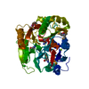
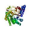
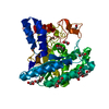
- PDB-3h7c: Crystal Structure of Arabidopsis thaliana Agmatine Deiminase from... -
+ Open data

- Basic information
| Entry | Database: PDB / ID: 3h7c | ||||||
|---|---|---|---|---|---|---|---|
| Title | Crystal Structure of Arabidopsis thaliana Agmatine Deiminase from Cell Free Expression | ||||||
 Components Components | Agmatine deiminase | ||||||
 Keywords Keywords | HYDROLASE / Agmatine / Structural Genomics / Protein Structure Initiative / PSI / Center for Eukaryotic Structural Genomics / CESG / N-carbamoylputrescine / Arginine decarboxylase pathway / Polyamine biosynthesis | ||||||
| Function / homology |  Function and homology information Function and homology informationagmatine deiminase / agmatine deiminase activity / : / polyamine biosynthetic process / protein-arginine deiminase activity / endoplasmic reticulum Similarity search - Function | ||||||
| Biological species |  | ||||||
| Method |  X-RAY DIFFRACTION / SYNCHROTRON / MAD / Resolution: 1.5 Å X-RAY DIFFRACTION / SYNCHROTRON / MAD / Resolution: 1.5 Å | ||||||
 Authors Authors | Burgie, E.S. / Bingman, C.A. / Phillips Jr., G.N. / Center for Eukaryotic Structural Genomics (CESG) | ||||||
 Citation Citation | Journal: To be Published Title: Structural Insights into the Catalytic Mechanism of Arabidopsis thaliana Agmatine Deiminase Authors: Burgie, E.S. / Bingman, C.A. / Phillips Jr., G.N. | ||||||
| History |
|
- Structure visualization
| Structure viewer | Molecule: MolmilJmol/JSmol |
|---|
- Downloads & links
-Download
| PDBx/mmCIF format | 3h7c.cif.gz | 101.6 KB | Display | PDBx/mmCIF format |
|---|---|---|---|---|
| PDB format | pdb3h7c.ent.gz | 77 KB | Display | PDB format |
| PDBx/mmJSON format | 3h7c.json.gz | Tree view | PDBx/mmJSON format | |
| Others |  Other downloads Other downloads |
-Validation report
| Arichive directory | https://data.pdbj.org/pub/pdb/validation_reports/h7/3h7cftp://data.pdbj.org/pub/pdb/validation_reports/h7/3h7c | HTTPS FTP |
|---|
-Related structure data


-Links
 PDBj
PDBj- Assembly
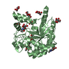
| Deposited unit | 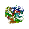
| ||||||||
|---|---|---|---|---|---|---|---|---|---|
| 1 |
| ||||||||
| Unit cell |
| ||||||||
| Details | biological unit is the same as asymmetric unit. |
-Components
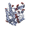
-Protein , 1 types, 1 molecules X
| #1: Protein | Mass: 43430.402 Da / Num. of mol.: 1 Source method: isolated from a genetically manipulated source Details: Cell free synthesis / Source: (gene. exp.) |
|---|
-Non-polymers , 7 types, 370 molecules 


| #2: Chemical |  Mass: 35.453 Da / Num. of mol.: 2 / Source method: obtained synthetically / Formula: Cl Mass: 35.453 Da / Num. of mol.: 2 / Source method: obtained synthetically / Formula: Cl#3: Chemical | ChemComp-MG / |  Mass: 24.305 Da / Num. of mol.: 1 / Source method: obtained synthetically / Formula: Mg Mass: 24.305 Da / Num. of mol.: 1 / Source method: obtained synthetically / Formula: Mg#4: Chemical |  Mass: 22.990 Da / Num. of mol.: 2 / Source method: obtained synthetically / Formula: Na Mass: 22.990 Da / Num. of mol.: 2 / Source method: obtained synthetically / Formula: Na#5: Chemical | ChemComp-K / |  Mass: 39.098 Da / Num. of mol.: 1 / Source method: obtained synthetically / Formula: K Mass: 39.098 Da / Num. of mol.: 1 / Source method: obtained synthetically / Formula: K#6: Chemical | ChemComp-211 / |  Mass: 149.188 Da / Num. of mol.: 1 / Source method: obtained synthetically / Formula: C6H15NO3 Mass: 149.188 Da / Num. of mol.: 1 / Source method: obtained synthetically / Formula: C6H15NO3#7: Chemical | ChemComp-1PE / |  Mass: 238.278 Da / Num. of mol.: 1 / Source method: obtained synthetically / Formula: C10H22O6 / Comment: precipitant*YM Mass: 238.278 Da / Num. of mol.: 1 / Source method: obtained synthetically / Formula: C10H22O6 / Comment: precipitant*YM#8: Water | ChemComp-HOH / | Mass: 18.015 Da / Num. of mol.: 362 / Source method: isolated from a natural source / Formula: H2O |
|---|
-Details
| Has protein modification | Y |
|---|---|
| Sequence details | AUTHORS STATE THAT THE SEQUENCE MATCHES GENBANK ENTRY AAO63405.1. THE DISCREPANCY BETWEEN AUTHOR'S ...AUTHORS STATE THAT THE SEQUENCE MATCHES GENBANK ENTRY AAO63405.1. THE DISCREPANC |
-Experimental details
-Experiment
| Experiment | Method: X-RAY DIFFRACTION / Number of used crystals: 1 |
|---|
- Sample preparation
| Crystal | Density Matthews: 2.51 Å3/Da / Density % sol: 50.97 % |
|---|---|
| Crystal grow | Temperature: 293 K / Method: vapor diffusion, hanging drop / pH: 7.5 Details: Protein solution- 10 mg/ml agmatine deiminase, 50 mM NaCl, 0.3 mM TCEP, 5 mM HEPES, pH 7.0; Precipitant solution- 34% Polyethylene glycol 2000, 200 mM KBr, 100 mM triethanolamine, pH 7.5; ...Details: Protein solution- 10 mg/ml agmatine deiminase, 50 mM NaCl, 0.3 mM TCEP, 5 mM HEPES, pH 7.0; Precipitant solution- 34% Polyethylene glycol 2000, 200 mM KBr, 100 mM triethanolamine, pH 7.5; Cryoprotectant- MiTeGen LV Cryo Oil, VAPOR DIFFUSION, HANGING DROP, temperature 293K |
-Data collection
| Diffraction | Mean temperature: 100 K | ||||||||||||
|---|---|---|---|---|---|---|---|---|---|---|---|---|---|
| Diffraction source | Source: SYNCHROTRON / Site: APS  / Beamline: 23-ID-D / Wavelength: 0.96350,0.97943,0.97957 / Beamline: 23-ID-D / Wavelength: 0.96350,0.97943,0.97957 | ||||||||||||
| Detector | Type: MARMOSAIC 300 mm CCD / Detector: CCD / Date: Apr 6, 2009 | ||||||||||||
| Radiation | Monochromator: Si(111) / Protocol: MAD / Monochromatic (M) / Laue (L): M / Scattering type: x-ray | ||||||||||||
| Radiation wavelength |
| ||||||||||||
| Reflection | Resolution: 1.5→50 Å / Num. all: 68398 / Num. obs: 68398 / % possible obs: 100 % / Redundancy: 7.5 % / Rmerge(I) obs: 0.098 / Net I/σ(I): 19.6 | ||||||||||||
| Reflection shell | Resolution: 1.5→1.53 Å / Redundancy: 6.7 % / Rmerge(I) obs: 0.444 / Mean I/σ(I) obs: 4.1 / Num. unique all: 3395 / % possible all: 100 |
-Phasing
| Phasing | Method: MAD |
|---|
- Processing
| Software |
| |||||||||||||||||||||||||||||||||||||||||||||||||||||||||||||||||
|---|---|---|---|---|---|---|---|---|---|---|---|---|---|---|---|---|---|---|---|---|---|---|---|---|---|---|---|---|---|---|---|---|---|---|---|---|---|---|---|---|---|---|---|---|---|---|---|---|---|---|---|---|---|---|---|---|---|---|---|---|---|---|---|---|---|---|
| Refinement | Method to determine structure: MAD / Resolution: 1.5→50 Å / Cor.coef. Fo:Fc: 0.968 / Cor.coef. Fo:Fc free: 0.955 / Occupancy max: 1 / Occupancy min: 0.2 / SU B: 0.884 / SU ML: 0.034 / Cross valid method: THROUGHOUT / σ(F): 0 / ESU R: 0.058 / ESU R Free: 0.062 / Stereochemistry target values: MAXIMUM LIKELIHOOD Details: HYDROGENS HAVE BEEN ADDED IN THE RIDING POSITIONS U VALUES : REFINED INDIVIDUALLY
| |||||||||||||||||||||||||||||||||||||||||||||||||||||||||||||||||
| Solvent computation | Ion probe radii: 0.8 Å / Shrinkage radii: 0.8 Å / VDW probe radii: 1.4 Å / Solvent model: BABINET MODEL WITH MASK | |||||||||||||||||||||||||||||||||||||||||||||||||||||||||||||||||
| Displacement parameters | Biso max: 71.57 Å2 / Biso mean: 14.686 Å2 / Biso min: 2 Å2
| |||||||||||||||||||||||||||||||||||||||||||||||||||||||||||||||||
| Refinement step | Cycle: LAST / Resolution: 1.5→50 Å
| |||||||||||||||||||||||||||||||||||||||||||||||||||||||||||||||||
| Refine LS restraints |
| |||||||||||||||||||||||||||||||||||||||||||||||||||||||||||||||||
| LS refinement shell | Resolution: 1.501→1.54 Å / Total num. of bins used: 20
|
