Mass: 18.015 Da / Num. of mol.: 327 / Source method: isolated from a natural source / Formula: H2O
-
Details
Has protein modification
Y
Sequence details
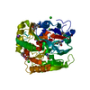
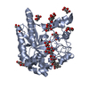
RESIDUE 90-134 ARE FROM HUMICOLA INSOLENS CEL6A. RESIDUE 135-308 AND 401-447 ARE FROM HYPOCREA ...RESIDUE 90-134 ARE FROM HUMICOLA INSOLENS CEL6A. RESIDUE 135-308 AND 401-447 ARE FROM HYPOCREA JECORINA CEL6A. RESIDUE 309-400 ARE FROM CHAETOMIUM THERMOPHILUM CEL6A.
-
Experimental details
-
Experiment
Experiment
Method: X-RAY DIFFRACTION / Number of used crystals: 1
-
Sample preparation
Crystal
Density Matthews: 2.62 Å3/Da / Density % sol: 53.14 %
Crystal grow
Method: vapor diffusion, sitting drop / pH: 6 / Details: pH 6.0, VAPOR DIFFUSION, SITTING DROP
Protocol: SINGLE WAVELENGTH / Monochromatic (M) / Laue (L): M / Scattering type: x-ray
Radiation wavelength
Wavelength: 1.033 Å / Relative weight: 1
Reflection
Resolution: 1.22→35.308 Å / Num. all: 106851 / Num. obs: 106851 / % possible obs: 87.3 % / Redundancy: 2.1 % / Rsym value: 0.027 / Net I/σ(I): 16.3
Reflection shell
Diffraction-ID: 1
Resolution (Å)
Redundancy (%)
Rmerge(I) obs
Mean I/σ(I) obs
Num. measured all
Num. unique all
Rsym value
% possible all
1.22-1.29
1.9
0.215
3.6
24424
12670
0.215
71.2
1.29-1.36
2
0.166
4.7
30030
14740
0.166
87.7
1.36-1.46
2
0.115
6.8
28965
14132
0.115
89.3
1.46-1.57
2.1
0.077
10.1
27819
13479
0.077
91.6
1.57-1.73
2.1
0.053
14.3
25769
12331
0.053
90.9
1.73-1.93
2.1
0.036
20.5
23921
11354
0.036
92.3
1.93-2.23
2.1
0.024
27.6
20655
9844
0.024
90.4
2.23-2.73
2.1
0.021
30.9
17581
8335
0.021
90.6
2.73-3.86
2.1
0.018
32.7
13586
6442
0.018
89.6
3.86-35.308
2.1
0.017
33.1
7344
3524
0.017
87.7
-
Phasing
Phasing
Method: molecular replacement
-
Processing
Software
Name
Version
Classification
NB
SCALA
3.3.16
datascaling
MOLREP
phasing
REFMAC
refinement
PDB_EXTRACT
3.11
dataextraction
Refinement
Method to determine structure: MOLECULAR REPLACEMENT / Resolution: 1.22→35.308 Å / Cor.coef. Fo:Fc: 0.975 / Cor.coef. Fo:Fc free: 0.97 / WRfactor Rfree: 0.1576 / WRfactor Rwork: 0.1424 / Occupancy max: 1 / Occupancy min: 0.5 / FOM work R set: 0.9289 / SU B: 0.875 / SU ML: 0.019 / SU R Cruickshank DPI: 0.0354 / SU Rfree: 0.0368 / Cross valid method: THROUGHOUT / σ(F): 0 / ESU R: 0.035 / ESU R Free: 0.037 / Stereochemistry target values: MAXIMUM LIKELIHOOD Details: HYDROGENS HAVE BEEN USED IF PRESENT IN THE INPUT U VALUES : RESIDUAL ONLY
Rfactor
Num. reflection
% reflection
Selection details
Rfree
0.1584
5345
5 %
RANDOM
Rwork
0.1426
-
-
-
obs
0.1434
106847
86.96 %
-
Solvent computation
Ion probe radii: 0.8 Å / Shrinkage radii: 0.8 Å / VDW probe radii: 1.2 Å / Solvent model: MASK
In the structure databanks used in Yorodumi, some data are registered as the other names, "COVID-19 virus" and "2019-nCoV". Here are the details of the virus and the list of structure data.
Jan 31, 2019. EMDB accession codes are about to change! (news from PDBe EMDB page)
EMDB accession codes are about to change! (news from PDBe EMDB page)
The allocation of 4 digits for EMDB accession codes will soon come to an end. Whilst these codes will remain in use, new EMDB accession codes will include an additional digit and will expand incrementally as the available range of codes is exhausted. The current 4-digit format prefixed with “EMD-” (i.e. EMD-XXXX) will advance to a 5-digit format (i.e. EMD-XXXXX), and so on. It is currently estimated that the 4-digit codes will be depleted around Spring 2019, at which point the 5-digit format will come into force.
The EM Navigator/Yorodumi systems omit the EMD- prefix.
Related info.:Q: What is EMD? / ID/Accession-code notation in Yorodumi/EM Navigator
Yorodumi is a browser for structure data from EMDB, PDB, SASBDB, etc.
This page is also the successor to EM Navigator detail page, and also detail information page/front-end page for Omokage search.
The word "yorodu" (or yorozu) is an old Japanese word meaning "ten thousand". "mi" (miru) is to see.
Related info.:EMDB / PDB / SASBDB / Comparison of 3 databanks / Yorodumi Search / Aug 31, 2016. New EM Navigator & Yorodumi / Yorodumi Papers / Jmol/JSmol / Function and homology information / Changes in new EM Navigator and Yorodumi
 Movie
Movie Controller


 Open data
 Basic information
 Components
 Keywords
 Function and homology information
 Humicola insolens (fungus)
 X-RAY DIFFRACTION /
  Authors
 Citation
 Structure visualization
 Downloads & links
 Other downloads
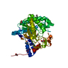


 PDBj
PDBj Assembly


 Mass: 59.044 Da / Num. of mol.: 2 / Source method: obtained synthetically / Formula: C2H3O2
 Mass: 62.068 Da / Num. of mol.: 10 / Source method: obtained synthetically / Formula: C2H6O2
 Mass: 194.226 Da / Num. of mol.: 1 / Source method: obtained synthetically / Formula: C8H18O5 / Comment: precipitant*YM
 Mass: 150.173 Da / Num. of mol.: 2 / Source method: obtained synthetically / Formula: C6H14O4
 Sample preparation
 / Beamline: BL12-2 / Wavelength: 1.033 Å
 Processing