- PDB-3h2b: Crystal structure of the SAM-dependent methyltransferase cg3271 f... -
+
Open data
ID or keywords:
Loading...
-
Basic information
Entry
Database: PDB / ID: 3h2b
Title
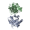
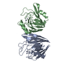
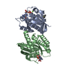
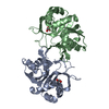
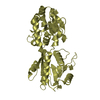
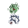
Crystal structure of the SAM-dependent methyltransferase cg3271 from Corynebacterium glutamicum in complex with S-adenosyl-L-homocysteine and pyrophosphate. Northeast Structural Genomics Consortium Target CgR113A
Resolution: 2→2.07 Å / Redundancy: 4.7 % / Rmerge(I) obs: 0.525 / Mean I/σ(I) obs: 2.62 / Num. unique all: 5044 / Rsym value: 0.49 / % possible all: 100
-
Processing
Software
Name
Version
Classification
NB
CNS
1.2
refinement
PDB_EXTRACT
3
dataextraction
MAR345dtb
datacollection
HKL-2000
datareduction
SCALEPACK
datascaling
SnB
phasing
SOLVE
phasing
RESOLVE
phasing
REFMAC
refinement
Refinement
Method to determine structure: SAD / Resolution: 2→19.97 Å / Rfactor Rfree error: 0.005 / Data cutoff high absF: 321463.094 / Data cutoff low absF: 0 / Isotropic thermal model: RESTRAINED / Cross valid method: THROUGHOUT / σ(F): 2 / σ(I): 2 / Stereochemistry target values: Engh & Huber Details: The Friedel pairs were used in phasing. Program XtalView has also been used in refinement.
In the structure databanks used in Yorodumi, some data are registered as the other names, "COVID-19 virus" and "2019-nCoV". Here are the details of the virus and the list of structure data.
Jan 31, 2019. EMDB accession codes are about to change! (news from PDBe EMDB page)
EMDB accession codes are about to change! (news from PDBe EMDB page)
The allocation of 4 digits for EMDB accession codes will soon come to an end. Whilst these codes will remain in use, new EMDB accession codes will include an additional digit and will expand incrementally as the available range of codes is exhausted. The current 4-digit format prefixed with “EMD-” (i.e. EMD-XXXX) will advance to a 5-digit format (i.e. EMD-XXXXX), and so on. It is currently estimated that the 4-digit codes will be depleted around Spring 2019, at which point the 5-digit format will come into force.
The EM Navigator/Yorodumi systems omit the EMD- prefix.
Related info.:Q: What is EMD? / ID/Accession-code notation in Yorodumi/EM Navigator
Yorodumi is a browser for structure data from EMDB, PDB, SASBDB, etc.
This page is also the successor to EM Navigator detail page, and also detail information page/front-end page for Omokage search.
The word "yorodu" (or yorozu) is an old Japanese word meaning "ten thousand". "mi" (miru) is to see.
Related info.:EMDB / PDB / SASBDB / Comparison of 3 databanks / Yorodumi Search / Aug 31, 2016. New EM Navigator & Yorodumi / Yorodumi Papers / Jmol/JSmol / Function and homology information / Changes in new EM Navigator and Yorodumi
 Movie
Movie Controller

 Yorodumi
 Open data
 Basic information
 Components
 Keywords
 Function and homology information
 Corynebacterium glutamicum ATCC 13032 (bacteria)
 X-RAY DIFFRACTION /
  Authors
 Citation
 Structure visualization
 Downloads & links
 Other downloads


 PDBj
PDBj Assembly

 Mass: 384.411 Da / Num. of mol.: 2 / Source method: obtained synthetically / Formula: C14H20N6O5S
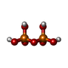
 Mass: 177.975 Da / Num. of mol.: 1 / Source method: obtained synthetically / Formula: H4O7P2
 Mass: 18.015 Da / Num. of mol.: 308 / Source method: isolated from a natural source / Formula: H2O
 Sample preparation
 / Beamline: X4C / Wavelength: 0.97869 Å
 Processing