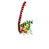
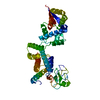
 Movie
Movie Controller

[English] 日本語
 Yorodumi
- PDB-3gyw: Crystal structure of nucleosome assembly protein from Plasmodium ... -
+ Open data

- Basic information
| Entry | Database: PDB / ID: 3gyw | ||||||
|---|---|---|---|---|---|---|---|
| Title | Crystal structure of nucleosome assembly protein from Plasmodium falciparum at 2.4 A resolution | ||||||
 Components Components | Nucleosome assembly protein 1, putative | ||||||
 Keywords Keywords | CHAPERONE / SIR / SIRAS / NUCLEOSOME ASSEMBLY PROTEIN / HISTONE RECOGNITION | ||||||
| Function / homology |  Function and homology information Function and homology informationnucleosome assembly / histone binding / chromatin binding / protein kinase binding / chromatin / nucleus / cytoplasm Similarity search - Function | ||||||
| Biological species |  | ||||||
| Method |  X-RAY DIFFRACTION / SIRAS / Resolution: 2.4 Å X-RAY DIFFRACTION / SIRAS / Resolution: 2.4 Å | ||||||
 Authors Authors | Yogavel, M. / Gill, J. / Sharma, A. | ||||||
 Citation Citation | Journal: Acta Crystallogr.,Sect.D / Year: 2009 Title: Iodide-SAD, SIR and SIRAS phasing for structure solution of a nucleosome assembly protein. Authors: Yogavel, M. / Gill, J. / Sharma, A. | ||||||
| History |
|
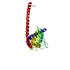
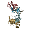
- Structure visualization
| Structure viewer | Molecule: MolmilJmol/JSmol |
|---|
- Downloads & links
-Download
| PDBx/mmCIF format | 3gyw.cif.gz | 51.3 KB | Display | PDBx/mmCIF format |
|---|---|---|---|---|
| PDB format | pdb3gyw.ent.gz | 35.8 KB | Display | PDB format |
| PDBx/mmJSON format | 3gyw.json.gz | Tree view | PDBx/mmJSON format | |
| Others |  Other downloads Other downloads |
-Validation report
| Arichive directory | https://data.pdbj.org/pub/pdb/validation_reports/gy/3gywftp://data.pdbj.org/pub/pdb/validation_reports/gy/3gyw | HTTPS FTP |
|---|
-Related structure data


-Links
 PDBj
PDBj
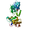
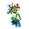
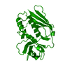
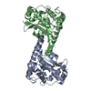
- Assembly
| Deposited unit | 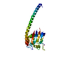
| ||||||||
|---|---|---|---|---|---|---|---|---|---|
| 1 | 
| ||||||||
| Unit cell |
|
-Components
| #1: Protein | Mass: 29667.742 Da / Num. of mol.: 1 / Fragment: residues 33-281 (UNP residues 45-293) Source method: isolated from a genetically manipulated source Source: (gene. exp.) Strain: 3D7 / Gene: pfl0185c / Plasmid: PET28A / Production host:  |
|---|---|
| #2: Water | ChemComp-HOH /  Mass: 18.015 Da / Num. of mol.: 43 / Source method: isolated from a natural source / Formula: H2O Mass: 18.015 Da / Num. of mol.: 43 / Source method: isolated from a natural source / Formula: H2O |
-Experimental details
-Experiment
| Experiment | Method: X-RAY DIFFRACTION / Number of used crystals: 1 |
|---|
- Sample preparation
| Crystal | Density Matthews: 1.9 Å3/Da / Density % sol: 35.19 % |
|---|---|
| Crystal grow | Temperature: 293 K / Method: vapor diffusion, hanging drop / pH: 7.6 Details: 20% PEG 3350, 0.2M magnesium chloride, pH 7.6, VAPOR DIFFUSION, HANGING DROP, temperature 293K |
-Data collection
| Diffraction | Mean temperature: 100 K |
|---|---|
| Diffraction source | Source: ROTATING ANODE / Type: RIGAKU MICROMAX-007 / Wavelength: 1.5418 Å |
| Detector | Type: MAR scanner 345 mm plate / Detector: IMAGE PLATE / Date: Nov 8, 2006 / Details: mirrors |
| Radiation | Monochromator: GRAPHITE / Protocol: SINGLE WAVELENGTH / Monochromatic (M) / Laue (L): M / Scattering type: x-ray |
| Radiation wavelength | Wavelength: 1.5418 Å / Relative weight: 1 |
| Reflection | Resolution: 2.4→50 Å / Num. all: 8943 / Num. obs: 8943 / % possible obs: 100 % / Observed criterion σ(F): 2 / Observed criterion σ(I): 2 / Redundancy: 2.4 % / Rmerge(I) obs: 0.04 / Net I/σ(I): 37.6 |
| Reflection shell | Resolution: 2.4→2.49 Å / Redundancy: 2.4 % / Rmerge(I) obs: 0.206 / Mean I/σ(I) obs: 8.1 / Num. unique all: 892 / % possible all: 100 |
- Processing
| Software |
| |||||||||||||||||||||||||
|---|---|---|---|---|---|---|---|---|---|---|---|---|---|---|---|---|---|---|---|---|---|---|---|---|---|---|
| Refinement | Method to determine structure: SIRAS / Resolution: 2.4→50 Å / σ(F): 2 / Stereochemistry target values: Engh & Huber
| |||||||||||||||||||||||||
| Refinement step | Cycle: LAST / Resolution: 2.4→50 Å
| |||||||||||||||||||||||||
| Refine LS restraints |
| |||||||||||||||||||||||||
| LS refinement shell | Resolution: 2.4→2.462 Å
|
