 Movie
Movie Controller


+ Open data

- Basic information
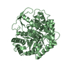
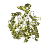
| Entry | Database: PDB / ID: 3guu | ||||||
|---|---|---|---|---|---|---|---|
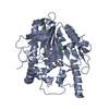
| Title | X-ray structure of Candida Antarctica lipase A | ||||||
 Components Components | Lipase A | ||||||
 Keywords Keywords | HYDROLASE / Candida / Lipase / Protein structure | ||||||
| Function / homology |  Function and homology information Function and homology informationtriacylglycerol lipase / triacylglycerol lipase activity / lipid catabolic process / extracellular region Similarity search - Function | ||||||
| Biological species |  Candida Antarctica (fungus) Candida Antarctica (fungus) | ||||||
| Method |  X-RAY DIFFRACTION / SYNCHROTRON / MOLECULAR REPLACEMENT / Resolution: 2.1 Å X-RAY DIFFRACTION / SYNCHROTRON / MOLECULAR REPLACEMENT / Resolution: 2.1 Å | ||||||
 Authors Authors | Brandt, A.-M. / Li, X.-G. / Nymalm-Rejstrom, Y. / Airenne, T. / Kanerva, L.T. / Salminen, T.A. | ||||||
 Citation Citation | Journal: To be Published Title: The crystal structure of Lipase A from Candida antarctica Authors: Brandt, A.-M. / Li, X.-G. / Nymalm-Rejstrom, Y. / Airenne, T. / Kanerva, L.T. / Salminen, T.A. | ||||||
| History |
|
- Structure visualization
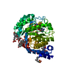
| Structure viewer | Molecule: MolmilJmol/JSmol |
|---|
- Downloads & links
-Download
| PDBx/mmCIF format | 3guu.cif.gz | 184.7 KB | Display | PDBx/mmCIF format |
|---|---|---|---|---|
| PDB format | pdb3guu.ent.gz | 145.2 KB | Display | PDB format |
| PDBx/mmJSON format | 3guu.json.gz | Tree view | PDBx/mmJSON format | |
| Others |  Other downloads Other downloads |
-Validation report
| Arichive directory | https://data.pdbj.org/pub/pdb/validation_reports/gu/3guuftp://data.pdbj.org/pub/pdb/validation_reports/gu/3guu | HTTPS FTP |
|---|
-Related structure data
| Related structure data |  2veoS S: Starting model for refinement |
|---|---|
| Similar structure data |


-Links
 PDBj
PDBj- Assembly
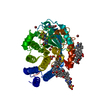
| Deposited unit | 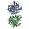
| ||||||||||||||||||
|---|---|---|---|---|---|---|---|---|---|---|---|---|---|---|---|---|---|---|---|
| 1 | 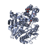
| ||||||||||||||||||
| 2 | 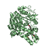
| ||||||||||||||||||
| Unit cell |
| ||||||||||||||||||
| Noncrystallographic symmetry (NCS) | NCS domain:
NCS domain segments: Component-ID: 1 / Ens-ID: 1 / Beg auth comp-ID: ALA / Beg label comp-ID: ALA / End auth comp-ID: PRO / End label comp-ID: PRO / Refine code: 5 / Auth seq-ID: 12 - 441 / Label seq-ID: 33 - 462
|
-Components
| #1: Protein | Mass: 49345.602 Da / Num. of mol.: 2 / Source method: isolated from a natural source / Details: Commerial enzyme Roche Chirazyme L-5 / Source: (natural) Candida Antarctica (fungus) / References: UniProt: W3VKA4*PLUS, triacylglycerol lipase#2: Chemical |   Mass: 238.278 Da / Num. of mol.: 2 / Source method: obtained synthetically / Formula: C10H22O6 / Comment: precipitant*YM Mass: 238.278 Da / Num. of mol.: 2 / Source method: obtained synthetically / Formula: C10H22O6 / Comment: precipitant*YM#3: Chemical | ChemComp-GOL / |   Mass: 92.094 Da / Num. of mol.: 1 / Source method: obtained synthetically / Formula: C3H8O3 Mass: 92.094 Da / Num. of mol.: 1 / Source method: obtained synthetically / Formula: C3H8O3#4: Chemical | ChemComp-SO4 / |   Mass: 96.063 Da / Num. of mol.: 1 / Source method: obtained synthetically / Formula: SO4 Mass: 96.063 Da / Num. of mol.: 1 / Source method: obtained synthetically / Formula: SO4#5: Water | ChemComp-HOH / |  Mass: 18.015 Da / Num. of mol.: 499 / Source method: isolated from a natural source / Formula: H2O Mass: 18.015 Da / Num. of mol.: 499 / Source method: isolated from a natural source / Formula: H2OHas protein modification | Y | Sequence details | THERE IS NO UNP REFERENCE SEQUENCE DATABASE FOR THIS PROTEIN AT THE TIME OF PROCESSING. THIS ...THERE IS NO UNP REFERENCE SEQUENCE DATABASE FOR THIS PROTEIN AT THE TIME OF PROCESSING | |
|---|
-Experimental details
-Experiment
| Experiment | Method: X-RAY DIFFRACTION / Number of used crystals: 1 |
|---|
- Sample preparation
| Crystal | Density Matthews: 3.23 Å3/Da / Density % sol: 61.89 % |
|---|---|
| Crystal grow | Temperature: 294 K / Method: vapor diffusion, hanging drop / pH: 8.5 Details: 1.5M Ammonium sulfate, 12% glycerol, 100mM TRIS-HCl pH 8.5 , VAPOR DIFFUSION, HANGING DROP, temperature 294.0K |
-Data collection
| Diffraction | Mean temperature: 100 K |
|---|---|
| Diffraction source | Source: SYNCHROTRON / Site: ESRF  / Beamline: ID14-2 / Wavelength: 0.933 Å / Beamline: ID14-2 / Wavelength: 0.933 Å |
| Detector | Type: ADSC QUANTUM 4 / Detector: CCD / Date: Dec 3, 2007 |
| Radiation | Protocol: SINGLE WAVELENGTH / Monochromatic (M) / Laue (L): M / Scattering type: x-ray |
| Radiation wavelength | Wavelength: 0.933 Å / Relative weight: 1 |
| Reflection | Resolution: 2.1→20 Å / Num. all: 88351 / Num. obs: 86764 / % possible obs: 98.2 % / Observed criterion σ(I): -3 / Redundancy: 6.5 % / Biso Wilson estimate: 20.9 Å2 / Rmerge(I) obs: 0.174 / Net I/σ(I): 9.64 |
| Reflection shell | Resolution: 2.1→2.3 Å / Redundancy: 6.3 % / Rmerge(I) obs: 0.433 / Mean I/σ(I) obs: 4.42 / Num. unique all: 17290 / Rsym value: 0.434 / % possible all: 96.4 |
- Processing
| Software |
| ||||||||||||||||||||||||||||||||||||||||||||||||||||||||||||||||||||||||||||||||||||||||||||||||||||||||||||||
|---|---|---|---|---|---|---|---|---|---|---|---|---|---|---|---|---|---|---|---|---|---|---|---|---|---|---|---|---|---|---|---|---|---|---|---|---|---|---|---|---|---|---|---|---|---|---|---|---|---|---|---|---|---|---|---|---|---|---|---|---|---|---|---|---|---|---|---|---|---|---|---|---|---|---|---|---|---|---|---|---|---|---|---|---|---|---|---|---|---|---|---|---|---|---|---|---|---|---|---|---|---|---|---|---|---|---|---|---|---|---|---|
| Refinement | Method to determine structure: MOLECULAR REPLACEMENT Starting model: PDB ENTRY 2VEO Resolution: 2.1→19.92 Å / Cor.coef. Fo:Fc: 0.929 / Cor.coef. Fo:Fc free: 0.892 / SU B: 4.309 / SU ML: 0.114 / Cross valid method: THROUGHOUT / ESU R: 0.166 / ESU R Free: 0.158 / Stereochemistry target values: MAXIMUM LIKELIHOOD / Details: HYDROGENS HAVE BEEN ADDED IN THE RIDING POSITIONS
| ||||||||||||||||||||||||||||||||||||||||||||||||||||||||||||||||||||||||||||||||||||||||||||||||||||||||||||||
| Solvent computation | Ion probe radii: 0.8 Å / Shrinkage radii: 0.8 Å / VDW probe radii: 1.2 Å / Solvent model: MASK | ||||||||||||||||||||||||||||||||||||||||||||||||||||||||||||||||||||||||||||||||||||||||||||||||||||||||||||||
| Displacement parameters | Biso mean: 14.292 Å2
| ||||||||||||||||||||||||||||||||||||||||||||||||||||||||||||||||||||||||||||||||||||||||||||||||||||||||||||||
| Refinement step | Cycle: LAST / Resolution: 2.1→19.92 Å
| ||||||||||||||||||||||||||||||||||||||||||||||||||||||||||||||||||||||||||||||||||||||||||||||||||||||||||||||
| Refine LS restraints |
| ||||||||||||||||||||||||||||||||||||||||||||||||||||||||||||||||||||||||||||||||||||||||||||||||||||||||||||||
| Refine LS restraints NCS | Dom-ID: 1 / Auth asym-ID: A / Ens-ID: 1 / Refine-ID: X-RAY DIFFRACTION
| ||||||||||||||||||||||||||||||||||||||||||||||||||||||||||||||||||||||||||||||||||||||||||||||||||||||||||||||
| LS refinement shell | Resolution: 2.1→2.154 Å / Total num. of bins used: 20
|
