 Movie
Movie Controller

[English] 日本語
 Yorodumi
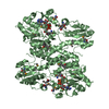
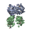
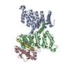
- PDB-3gnt: Crystal Structure of the Staphylococcus aureus Enoyl-Acyl Carrier... -
+ Open data

- Basic information
| Entry | Database: PDB / ID: 3gnt | ||||||
|---|---|---|---|---|---|---|---|
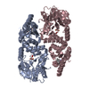
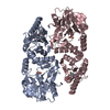
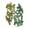
| Title | Crystal Structure of the Staphylococcus aureus Enoyl-Acyl Carrier Protein Reductase (FabI) in apo form (two molecules in AU) | ||||||
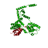
 Components Components | Enoyl-[acyl-carrier-protein] reductase [NADH] | ||||||
 Keywords Keywords | OXIDOREDUCTASE / Enoyl reductase / Rossmann fold | ||||||
| Function / homology |  Function and homology information Function and homology informationenoyl-[acyl-carrier-protein] reductase (NADPH, Re-specific) / enoyl-[acyl-carrier-protein] reductase (NADPH) activity / fatty acid elongation / enoyl-[acyl-carrier-protein] reductase (NADH) activity / NADP binding / identical protein binding Similarity search - Function | ||||||
| Biological species |   Staphylococcus aureus (bacteria) Staphylococcus aureus (bacteria) | ||||||
| Method |  X-RAY DIFFRACTION / SYNCHROTRON / MOLECULAR REPLACEMENT / Resolution: 2.75 Å X-RAY DIFFRACTION / SYNCHROTRON / MOLECULAR REPLACEMENT / Resolution: 2.75 Å | ||||||
 Authors Authors | Priyadarshi, A. / Hwang, K.Y. | ||||||
 Citation Citation | Journal: Proteins / Year: 2010 Title: Structural insights into Staphylococcus aureus enoyl-ACP reductase (FabI), in complex with NADP and triclosan. Authors: Priyadarshi, A. / Kim, E.E. / Hwang, K.Y. | ||||||
| History |
|
- Structure visualization
| Structure viewer | Molecule: MolmilJmol/JSmol |
|---|
- Downloads & links
-Download
| PDBx/mmCIF format | 3gnt.cif.gz | 91.9 KB | Display | PDBx/mmCIF format |
|---|---|---|---|---|
| PDB format | pdb3gnt.ent.gz | 70.5 KB | Display | PDB format |
| PDBx/mmJSON format | 3gnt.json.gz | Tree view | PDBx/mmJSON format | |
| Others |  Other downloads Other downloads |
-Validation report
| Arichive directory | https://data.pdbj.org/pub/pdb/validation_reports/gn/3gntftp://data.pdbj.org/pub/pdb/validation_reports/gn/3gnt | HTTPS FTP |
|---|
-Related structure data
| Related structure data | 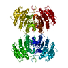 3gnsC 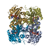 3gr6C 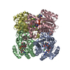 2pd3S C: citing same article ( S: Starting model for refinement |
|---|---|
| Similar structure data |


-Links
 PDBj
PDBj

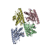
- Assembly
| Deposited unit | 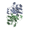
| ||||||||
|---|---|---|---|---|---|---|---|---|---|
| 1 | 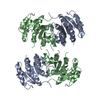
| ||||||||
| 2 |
| ||||||||
| Unit cell |
|
-Components
| #1: Protein | Mass: 28024.834 Da / Num. of mol.: 2 Source method: isolated from a genetically manipulated source Source: (gene. exp.) Staphylococcus aureus (bacteria) / Strain: MRSA252 / Gene: fabI / Plasmid: pET28a / Production host: References: UniProt: Q6GI75, enoyl-[acyl-carrier-protein] reductase (NADH) |
|---|
-Experimental details
-Experiment
| Experiment | Method: X-RAY DIFFRACTION / Number of used crystals: 1 |
|---|
- Sample preparation
| Crystal | Density Matthews: 2.35 Å3/Da / Density % sol: 47.72 % |
|---|---|
| Crystal grow | Temperature: 295 K / Method: vapor diffusion, hanging drop / pH: 8 Details: 15% PEG 400, 0.1M Tris pH 8.0, 4% glycerol, VAPOR DIFFUSION, HANGING DROP, temperature 295K |
-Data collection
| Diffraction | Mean temperature: 100 K |
|---|---|
| Diffraction source | Source: SYNCHROTRON / Site: PAL/PLS  / Beamline: 6C1 / Wavelength: 1 Å / Beamline: 6C1 / Wavelength: 1 Å |
| Detector | Type: ADSC QUANTUM 270 / Detector: CCD / Date: May 8, 2008 / Details: mirrors |
| Radiation | Monochromator: GRAPHITE / Protocol: SINGLE WAVELENGTH / Monochromatic (M) / Laue (L): M / Scattering type: x-ray |
| Radiation wavelength | Wavelength: 1 Å / Relative weight: 1 |
| Reflection | Resolution: 2.75→50 Å / Num. all: 11436 / Num. obs: 10886 / % possible obs: 96 % / Observed criterion σ(F): 2 / Observed criterion σ(I): 2 / Redundancy: 4.2 % / Biso Wilson estimate: 36 Å2 / Rmerge(I) obs: 0.09 / Rsym value: 0.17 / Net I/σ(I): 12.7 |
| Reflection shell | Resolution: 2.75→2.85 Å / Redundancy: 2.1 % / Rmerge(I) obs: 0.18 / Mean I/σ(I) obs: 2.4 / Num. unique all: 635 / Rsym value: 0.17 / % possible all: 47 |
- Processing
| Software |
| |||||||||||||||||||||||||||||||||||||||||||||||||||||||||||||||||||||||||||||||||||||
|---|---|---|---|---|---|---|---|---|---|---|---|---|---|---|---|---|---|---|---|---|---|---|---|---|---|---|---|---|---|---|---|---|---|---|---|---|---|---|---|---|---|---|---|---|---|---|---|---|---|---|---|---|---|---|---|---|---|---|---|---|---|---|---|---|---|---|---|---|---|---|---|---|---|---|---|---|---|---|---|---|---|---|---|---|---|---|
| Refinement | Method to determine structure: MOLECULAR REPLACEMENT Starting model: 2PD3 Resolution: 2.75→34.96 Å / Cor.coef. Fo:Fc: 0.879 / Cor.coef. Fo:Fc free: 0.869 / SU B: 13.349 / SU ML: 0.278 / Cross valid method: THROUGHOUT / σ(F): 0 / σ(I): 0 / ESU R Free: 0.444 / Stereochemistry target values: MAXIMUM LIKELIHOOD / Details: HYDROGENS HAVE BEEN ADDED IN THE RIDING POSITIONS
| |||||||||||||||||||||||||||||||||||||||||||||||||||||||||||||||||||||||||||||||||||||
| Solvent computation | Ion probe radii: 0.8 Å / Shrinkage radii: 0.8 Å / VDW probe radii: 1.4 Å / Solvent model: MASK | |||||||||||||||||||||||||||||||||||||||||||||||||||||||||||||||||||||||||||||||||||||
| Displacement parameters | Biso mean: 36.972 Å2
| |||||||||||||||||||||||||||||||||||||||||||||||||||||||||||||||||||||||||||||||||||||
| Refinement step | Cycle: LAST / Resolution: 2.75→34.96 Å
| |||||||||||||||||||||||||||||||||||||||||||||||||||||||||||||||||||||||||||||||||||||
| Refine LS restraints |
| |||||||||||||||||||||||||||||||||||||||||||||||||||||||||||||||||||||||||||||||||||||
| LS refinement shell | Resolution: 2.752→2.823 Å / Rfactor Rfree error: 0.015 / Total num. of bins used: 20
|
