 Movie
Movie Controller

[English] 日本語
 Yorodumi
- PDB-3gmg: Crystal structure of an uncharacterized conserved protein from My... -
+ Open data

- Basic information
| Entry | Database: PDB / ID: 3gmg | ||||||
|---|---|---|---|---|---|---|---|
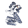
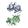
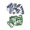
| Title | Crystal structure of an uncharacterized conserved protein from Mycobacterium tuberculosis | ||||||
 Components Components | Uncharacterized protein Rv1825/MT1873 | ||||||
 Keywords Keywords | STRUCTURAL GENOMICS / UNKNOWN FUNCTION / Cell membrane / Membrane / Transmembrane / PSI-2 / Protein Structure Initiative / New York SGX Research Center for Structural Genomics / NYSGXRC | ||||||
| Function / homology |  Function and homology information Function and homology information | ||||||
| Biological species |   Mycobacterium tuberculosis (bacteria) Mycobacterium tuberculosis (bacteria) | ||||||
| Method |  X-RAY DIFFRACTION / SYNCHROTRON / SAD / Resolution: 1.5 Å X-RAY DIFFRACTION / SYNCHROTRON / SAD / Resolution: 1.5 Å | ||||||
 Authors Authors | Bonanno, J.B. / Rutter, M. / Bain, K.T. / Chang, S. / Ozyurt, S. / Wasserman, S. / Sauder, J.M. / Burley, S.K. / Almo, S.C. / New York SGX Research Center for Structural Genomics (NYSGXRC) | ||||||
 Citation Citation | Journal: To be Published Title: Crystal structure of an uncharacterized conserved protein from Mycobacterium tuberculosis Authors: Bonanno, J.B. / Rutter, M. / Bain, K.T. / Chang, S. / Ozyurt, S. / Wasserman, S. / Sauder, J.M. / Burley, S.K. / Almo, S.C. | ||||||
| History |
|
- Structure visualization
| Structure viewer | Molecule: MolmilJmol/JSmol |
|---|
- Downloads & links
-Download
| PDBx/mmCIF format | 3gmg.cif.gz | 70.1 KB | Display | PDBx/mmCIF format |
|---|---|---|---|---|
| PDB format | pdb3gmg.ent.gz | 51.8 KB | Display | PDB format |
| PDBx/mmJSON format | 3gmg.json.gz | Tree view | PDBx/mmJSON format | |
| Others |  Other downloads Other downloads |
-Validation report
| Summary document | 3gmg_validation.pdf.gz | 428.3 KB | Display | wwPDB validaton report |
|---|---|---|---|---|
| Full document | 3gmg_full_validation.pdf.gz | 430.8 KB | Display | |
| Data in XML | 3gmg_validation.xml.gz | 14.9 KB | Display | |
| Data in CIF | 3gmg_validation.cif.gz | 21.6 KB | Display | |
| Arichive directory | https://data.pdbj.org/pub/pdb/validation_reports/gm/3gmgftp://data.pdbj.org/pub/pdb/validation_reports/gm/3gmg | HTTPS FTP |
-Related structure data
| Similar structure data | |
|---|---|
| Other databases |


-Links
 PDBj
PDBj- Assembly
| Deposited unit | 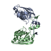
| ||||||||
|---|---|---|---|---|---|---|---|---|---|
| 1 |
| ||||||||
| Unit cell |
| ||||||||
| Details | AUTHORS STATE THAT THE DIMERIC ASSEMBLY OF THE BIOLOGICAL UNIT THAT IS SHOWN IN REMARK 350 IS PUTATIVE. |
-Components
| #1: Protein | Mass: 17633.984 Da / Num. of mol.: 2 / Fragment: UNP residues 134-292 Source method: isolated from a genetically manipulated source Source: (gene. exp.) Mycobacterium tuberculosis (bacteria) / Gene: Rv1825, MT1873, MTCY1A11.18c / Plasmid: modified pET26 / Production host: #2: Water | ChemComp-HOH / |  Mass: 18.015 Da / Num. of mol.: 256 / Source method: isolated from a natural source / Formula: H2O Mass: 18.015 Da / Num. of mol.: 256 / Source method: isolated from a natural source / Formula: H2O |
|---|
-Experimental details
-Experiment
| Experiment | Method: X-RAY DIFFRACTION / Number of used crystals: 1 |
|---|
- Sample preparation
| Crystal | Density Matthews: 2.19 Å3/Da / Density % sol: 43.89 % |
|---|---|
| Crystal grow | Temperature: 294 K / Method: vapor diffusion / pH: 6.5 Details: 100mM Bis-Tris pH 6.5, 25% PEG 3350, 200mM Sodium chloride, VAPOR DIFFUSION, temperature 294K |
-Data collection
| Diffraction | Mean temperature: 100 K |
|---|---|
| Diffraction source | Source: SYNCHROTRON / Site: APS  / Beamline: 31-ID / Wavelength: 0.97958 Å / Beamline: 31-ID / Wavelength: 0.97958 Å |
| Detector | Type: MAR CCD 165 mm / Detector: CCD / Date: Mar 13, 2009 |
| Radiation | Monochromator: diamond / Protocol: SINGLE WAVELENGTH / Monochromatic (M) / Laue (L): M / Scattering type: x-ray |
| Radiation wavelength | Wavelength: 0.97958 Å / Relative weight: 1 |
| Reflection | Resolution: 1.5→51.723 Å / Num. all: 50353 / Num. obs: 50353 / % possible obs: 99.9 % / Observed criterion σ(F): 0 / Observed criterion σ(I): 0 / Redundancy: 14.6 % / Biso Wilson estimate: 15.2 Å2 / Rmerge(I) obs: 0.081 / Rsym value: 0.081 / Net I/σ(I): 25.2 |
| Reflection shell | Resolution: 1.5→1.58 Å / Redundancy: 14.6 % / Rmerge(I) obs: 0.289 / Mean I/σ(I) obs: 7.8 / Num. unique all: 7254 / Rsym value: 0.289 / % possible all: 100 |
- Processing
| Software |
| |||||||||||||||||||||||||||||||||||||||||||||||||||||||||||||||||||||||||||||||||||||
|---|---|---|---|---|---|---|---|---|---|---|---|---|---|---|---|---|---|---|---|---|---|---|---|---|---|---|---|---|---|---|---|---|---|---|---|---|---|---|---|---|---|---|---|---|---|---|---|---|---|---|---|---|---|---|---|---|---|---|---|---|---|---|---|---|---|---|---|---|---|---|---|---|---|---|---|---|---|---|---|---|---|---|---|---|---|---|
| Refinement | Method to determine structure: SAD / Resolution: 1.5→18 Å / Cor.coef. Fo:Fc: 0.957 / Cor.coef. Fo:Fc free: 0.942 / WRfactor Rfree: 0.218 / WRfactor Rwork: 0.19 / Occupancy max: 1 / Occupancy min: 0.5 / FOM work R set: 0.886 / SU B: 1.035 / SU ML: 0.04 / SU R Cruickshank DPI: 0.069 / SU Rfree: 0.071 / Cross valid method: THROUGHOUT / ESU R: 0.069 / ESU R Free: 0.071 / Stereochemistry target values: MAXIMUM LIKELIHOOD / Details: HYDROGENS HAVE BEEN ADDED IN THE RIDING POSITIONS
| |||||||||||||||||||||||||||||||||||||||||||||||||||||||||||||||||||||||||||||||||||||
| Solvent computation | Ion probe radii: 0.8 Å / Shrinkage radii: 0.8 Å / VDW probe radii: 1.2 Å / Solvent model: BABINET MODEL WITH MASK | |||||||||||||||||||||||||||||||||||||||||||||||||||||||||||||||||||||||||||||||||||||
| Displacement parameters | Biso max: 47.93 Å2 / Biso mean: 18.623 Å2 / Biso min: 8.22 Å2
| |||||||||||||||||||||||||||||||||||||||||||||||||||||||||||||||||||||||||||||||||||||
| Refinement step | Cycle: LAST / Resolution: 1.5→18 Å
| |||||||||||||||||||||||||||||||||||||||||||||||||||||||||||||||||||||||||||||||||||||
| Refine LS restraints |
| |||||||||||||||||||||||||||||||||||||||||||||||||||||||||||||||||||||||||||||||||||||
| LS refinement shell | Resolution: 1.5→1.539 Å / Total num. of bins used: 20
|
