 Movie
Movie Controller


+ Open data

- Basic information
| Entry | Database: PDB / ID: 3gm7 | ||||||||||||||||||||
|---|---|---|---|---|---|---|---|---|---|---|---|---|---|---|---|---|---|---|---|---|---|
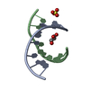
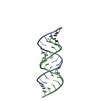
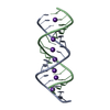
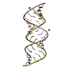
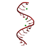
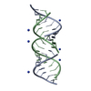
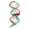
| Title | 1.58 A resolution X-ray structure of (CUG)6 | ||||||||||||||||||||
 Components Components | 5'-R(* Keywords KeywordsRNA / stretched U-U wobble / myotonic dystrophy / CUG repeats | Function / homology | RNA / RNA (> 10) |  Function and homology information Function and homology informationMethod |  X-RAY DIFFRACTION / PDB entry 1ZEV / Resolution: 1.58 Å X-RAY DIFFRACTION / PDB entry 1ZEV / Resolution: 1.58 Å  Authors AuthorsKiliszek, A. / Kierzek, R. / Krzyzosiak, W.J. / Rypniewski, W. |  CitationJournal: Proc.Natl.Acad.Sci.USA / Year: 2005 CitationJournal: Proc.Natl.Acad.Sci.USA / Year: 2005Title: The structural basis of myotonic dystrophy from the crystal structure of CUG repeats. Authors: Mooers, B.H. / Logue, J.S. / Berglund, J.A. History |
Remark 0 | THIS ENTRY (3GM7) REFLECTS AN ALTERNATIVE MODELING OF THE ORIGINAL STRUCTURE (PDB ID 1ZEV) ...THIS ENTRY (3GM7) REFLECTS AN ALTERNATIVE MODELING OF THE ORIGINAL STRUCTURE (PDB ID 1ZEV) DETERMINED BY THE AUTHORS: B.H. MOOERS, J.S. LOGUE, J.A. BERGLUND | |
- Structure visualization
| Structure viewer | Molecule: MolmilJmol/JSmol |
|---|
- Downloads & links
-Download
| PDBx/mmCIF format | 3gm7.cif.gz | 50.5 KB | Display | PDBx/mmCIF format |
|---|---|---|---|---|
| PDB format | pdb3gm7.ent.gz | 37.8 KB | Display | PDB format |
| PDBx/mmJSON format | 3gm7.json.gz | Tree view | PDBx/mmJSON format | |
| Others |  Other downloads Other downloads |
-Validation report
| Arichive directory | https://data.pdbj.org/pub/pdb/validation_reports/gm/3gm7ftp://data.pdbj.org/pub/pdb/validation_reports/gm/3gm7 | HTTPS FTP |
|---|
-Related structure data


-Links
 PDBj
PDBj


- Assembly
| Deposited unit | 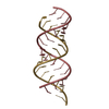
| ||||||||||||||||||||||||
|---|---|---|---|---|---|---|---|---|---|---|---|---|---|---|---|---|---|---|---|---|---|---|---|---|---|
| 1 |
| ||||||||||||||||||||||||
| Unit cell |
| ||||||||||||||||||||||||
| Components on special symmetry positions |
|
-Components
| #1: RNA chain | Mass: 5694.363 Da / Num. of mol.: 2 / Source method: obtained synthetically Details: Synthetic mRNA with the sequence of the part of human mRNA #2: Water | ChemComp-HOH / |  Mass: 18.015 Da / Num. of mol.: 52 / Source method: isolated from a natural source / Formula: H2O Mass: 18.015 Da / Num. of mol.: 52 / Source method: isolated from a natural source / Formula: H2O |
|---|
-Experimental details
-Experiment
| Experiment | Method: X-RAY DIFFRACTION / Number of used crystals: 1 |
|---|
- Sample preparation
| Crystal | Density Matthews: 1.83 Å3/Da / Density % sol: 32.61 % | ||||||||||||||||||||||||||||||||||||
|---|---|---|---|---|---|---|---|---|---|---|---|---|---|---|---|---|---|---|---|---|---|---|---|---|---|---|---|---|---|---|---|---|---|---|---|---|---|
| Crystal grow | Temperature: 277 K / Method: vapor diffusion, hanging drop / pH: 7 Details: MOPS, NaCl, MgCl2, MPD, pH 7.0, VAPOR DIFFUSION, HANGING DROP, temperature 277K | ||||||||||||||||||||||||||||||||||||
| Components of the solutions |
|
-Data collection
| Radiation | Scattering type: x-ray |
|---|---|
| Radiation wavelength | Relative weight: 1 |
- Processing
| Software | Name: SHELXL-97 / Classification: refinement | ||||||||||||||||||||||||
|---|---|---|---|---|---|---|---|---|---|---|---|---|---|---|---|---|---|---|---|---|---|---|---|---|---|
| Refinement | Method to determine structure: PDB entry 1ZEV / Resolution: 1.58→19.6 Å / Cross valid method: THROUGHOUT Details: AUTHORS STATE THAT ALL THE ATOMS THAT ARE LISTED IN REMARK 500 FIT WELL THE ELECTRON DENSITY.
| ||||||||||||||||||||||||
| Displacement parameters | Biso mean: 33.8 Å2 | ||||||||||||||||||||||||
| Refinement step | Cycle: LAST / Resolution: 1.58→19.6 Å
| ||||||||||||||||||||||||
| Refine LS restraints |
| ||||||||||||||||||||||||
| LS refinement shell | Resolution: 1.58→1.64 Å
|
