 Movie
Movie Controller

[English] 日本語
 Yorodumi
- PDB-3g9w: Crystal Structure of Talin2 F2-F3 in Complex with the Integrin Be... -
+ Open data

- Basic information
| Entry | Database: PDB / ID: 3g9w | ||||||
|---|---|---|---|---|---|---|---|
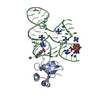
| Title | Crystal Structure of Talin2 F2-F3 in Complex with the Integrin Beta1D Cytoplasmic Tail | ||||||
 Components Components |
| ||||||
 Keywords Keywords | CELL ADHESION / protein-protein complex / PH domain superfold / PTB domain / helical bundle / intrinsically unstructured | ||||||
| Function / homology |  Function and homology information Function and homology informationpresynaptic actin cytoskeleton organization / integrin alpha8-beta1 complex / myoblast fate specification / integrin alpha3-beta1 complex / integrin alpha6-beta1 complex / integrin alpha7-beta1 complex / integrin alpha10-beta1 complex / integrin alpha11-beta1 complex / positive regulation of glutamate uptake involved in transmission of nerve impulse / cardiac cell fate specification ...presynaptic actin cytoskeleton organization / integrin alpha8-beta1 complex / myoblast fate specification / integrin alpha3-beta1 complex / integrin alpha6-beta1 complex / integrin alpha7-beta1 complex / integrin alpha10-beta1 complex / integrin alpha11-beta1 complex / positive regulation of glutamate uptake involved in transmission of nerve impulse / cardiac cell fate specification / integrin alpha5-beta1 complex / integrin alpha9-beta1 complex / regulation of collagen catabolic process / integrin alpha1-beta1 complex / integrin binding involved in cell-matrix adhesion / integrin alpha4-beta1 complex / cell adhesion receptor activity / collagen binding involved in cell-matrix adhesion / integrin alpha2-beta1 complex / Localization of the PINCH-ILK-PARVIN complex to focal adhesions / cell-cell adhesion mediated by integrin / formation of radial glial scaffolds / Other semaphorin interactions / Formation of the ureteric bud / cerebellar climbing fiber to Purkinje cell synapse / CD40 signaling pathway / calcium-independent cell-matrix adhesion / positive regulation of fibroblast growth factor receptor signaling pathway / reactive gliosis / myelin sheath abaxonal region / integrin alphav-beta1 complex / fascia adherens / basement membrane organization / CHL1 interactions / cardiac muscle cell myoblast differentiation / regulation of synapse pruning / Fibronectin matrix formation / MET interacts with TNS proteins / Laminin interactions / Developmental Lineage of Mammary Stem Cells / primordial germ cell migration / Platelet Adhesion to exposed collagen / leukocyte tethering or rolling / myoblast fusion / positive regulation of vascular endothelial growth factor signaling pathway / cell migration involved in sprouting angiogenesis / axon extension / Elastic fibre formation / cardiac muscle cell differentiation / myoblast differentiation / mesodermal cell differentiation / central nervous system neuron differentiation / Differentiation of Keratinocytes in Interfollicular Epidermis in Mammalian Skin / cell projection organization / wound healing, spreading of epidermal cells / positive regulation of fibroblast migration / negative regulation of Rho protein signal transduction / integrin complex / regulation of spontaneous synaptic transmission / cell adhesion mediated by integrin / Molecules associated with elastic fibres / MET activates PTK2 signaling / heterotypic cell-cell adhesion / Basigin interactions / lamellipodium assembly / negative regulation of vasoconstriction / sarcomere organization / muscle organ development / Mechanical load activates signaling by PIEZO1 and integrins in osteocytes / leukocyte cell-cell adhesion / Syndecan interactions / dendrite morphogenesis / positive regulation of neuroblast proliferation / negative regulation of neuron differentiation / positive regulation of wound healing / response to muscle activity / cell-substrate adhesion / establishment of mitotic spindle orientation / maintenance of blood-brain barrier / homophilic cell-cell adhesion / Developmental Lineage of Mammary Gland Myoepithelial Cells / cleavage furrow / TGF-beta receptor signaling activates SMADs / fibronectin binding / negative regulation of anoikis / neuroblast proliferation / RHOG GTPase cycle / intercalated disc / positive regulation of GTPase activity / RAC3 GTPase cycle / RAC2 GTPase cycle / ECM proteoglycans / Integrin cell surface interactions / cellular response to low-density lipoprotein particle stimulus / glial cell projection / cellular defense response / phagocytosis / coreceptor activity / ruffle / RAC1 GTPase cycle Similarity search - Function | ||||||
| Biological species |   Homo sapiens (human) Homo sapiens (human) | ||||||
| Method |  X-RAY DIFFRACTION / SYNCHROTRON / MOLECULAR REPLACEMENT / Resolution: 2.165 Å X-RAY DIFFRACTION / SYNCHROTRON / MOLECULAR REPLACEMENT / Resolution: 2.165 Å | ||||||
 Authors Authors | Anthis, N.J. / Wegener, K.L. / Ye, F. / Kim, C. / Lowe, E.D. / Vakonakis, I. / Bate, N. / Critchley, D.R. / Ginsberg, M.H. / Campbell, I.D. | ||||||
 Citation Citation | Journal: Embo J. / Year: 2009 Title: The structure of an integrin/talin complex reveals the basis of inside-out signal transduction Authors: Anthis, N.J. / Wegener, K.L. / Ye, F. / Kim, C. / Goult, B.T. / Lowe, E.D. / Vakonakis, I. / Bate, N. / Critchley, D.R. / Ginsberg, M.H. / Campbell, I.D. | ||||||
| History |
|
- Structure visualization
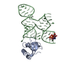
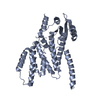
| Structure viewer | Molecule: MolmilJmol/JSmol |
|---|
- Downloads & links
-Download
| PDBx/mmCIF format | 3g9w.cif.gz | 226.9 KB | Display | PDBx/mmCIF format |
|---|---|---|---|---|
| PDB format | pdb3g9w.ent.gz | 182.3 KB | Display | PDB format |
| PDBx/mmJSON format | 3g9w.json.gz | Tree view | PDBx/mmJSON format | |
| Others |  Other downloads Other downloads |
-Validation report
| Arichive directory | https://data.pdbj.org/pub/pdb/validation_reports/g9/3g9wftp://data.pdbj.org/pub/pdb/validation_reports/g9/3g9w | HTTPS FTP |
|---|
-Related structure data
| Related structure data | 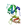 1mixS  1mk7S  1mk9S S: Starting model for refinement |
|---|---|
| Similar structure data | |
| Other databases |
|

-Links
 PDBj
PDBj


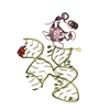
- Assembly
| Deposited unit | 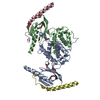
| ||||||||
|---|---|---|---|---|---|---|---|---|---|
| 1 | 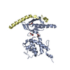
| ||||||||
| 2 | 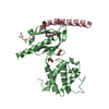
| ||||||||
| Unit cell |
|
-Components
| #1: Protein | Mass: 25762.516 Da / Num. of mol.: 2 / Fragment: F2-F3 domain Source method: isolated from a genetically manipulated source Source: (gene. exp.)  #2: Protein | Mass: 6190.161 Da / Num. of mol.: 2 / Fragment: Cytoplasmic Tail Source method: isolated from a genetically manipulated source Source: (gene. exp.) Homo sapiens (human) / Gene: ITGB1 / Plasmid: pET16b / Production host: #3: Chemical | ChemComp-GOL /   Mass: 92.094 Da / Num. of mol.: 6 / Source method: obtained synthetically / Formula: C3H8O3 Mass: 92.094 Da / Num. of mol.: 6 / Source method: obtained synthetically / Formula: C3H8O3#4: Chemical |   Mass: 106.120 Da / Num. of mol.: 2 / Source method: obtained synthetically / Formula: C4H10O3 Mass: 106.120 Da / Num. of mol.: 2 / Source method: obtained synthetically / Formula: C4H10O3#5: Water | ChemComp-HOH / |  Mass: 18.015 Da / Num. of mol.: 381 / Source method: isolated from a natural source / Formula: H2O Mass: 18.015 Da / Num. of mol.: 381 / Source method: isolated from a natural source / Formula: H2OSequence details | THE SEQUENCE OF CHAIN C, D IS ISOFORM BETA-1D AS LISTED IN UNP ENTRY, P05556-5 | |
|---|
-Experimental details
-Experiment
| Experiment | Method: X-RAY DIFFRACTION / Number of used crystals: 1 |
|---|
- Sample preparation
| Crystal | Density Matthews: 2.99 Å3/Da / Density % sol: 58.82 % |
|---|---|
| Crystal grow | Temperature: 277 K / Method: vapor diffusion, sitting drop / pH: 7 Details: 0.1M ammonium acetate, 0.02M magnesium chloride, 0.05M HEPES, 5% PEG 8k, pH 7.0, VAPOR DIFFUSION, SITTING DROP, temperature 277K |
-Data collection
| Diffraction | Mean temperature: 100 K |
|---|---|
| Diffraction source | Source: SYNCHROTRON / Site: ESRF  / Beamline: ID23-2 / Wavelength: 0.8726 Å / Beamline: ID23-2 / Wavelength: 0.8726 Å |
| Detector | Type: MARMOSAIC 225 mm CCD / Detector: CCD / Date: Jun 26, 2008 Details: Monochromator (horizontally side diffracting Silicon 111 crystal) |
| Radiation | Monochromator: horizontally diffracting monochromator / Protocol: SINGLE WAVELENGTH / Monochromatic (M) / Laue (L): M / Scattering type: x-ray |
| Radiation wavelength | Wavelength: 0.8726 Å / Relative weight: 1 |
| Reflection | Resolution: 2.165→41.95 Å / Num. all: 41528 / Num. obs: 41362 / % possible obs: 99.6 % / Observed criterion σ(F): 2 / Observed criterion σ(I): 2 / Redundancy: 3.7 % / Biso Wilson estimate: 25.312 Å2 / Rmerge(I) obs: 0.114 / Rsym value: 0.114 / Net I/σ(I): 8.3 |
| Reflection shell | Resolution: 2.165→2.28 Å / Redundancy: 3.7 % / Rmerge(I) obs: 0.338 / Mean I/σ(I) obs: 3.5 / Num. unique all: 5982 / Rsym value: 0.338 / % possible all: 99.9 |
- Processing
| Software |
| |||||||||||||||||||||||||||||||||||||||||||||||||||||||||||||||||||||||||||||||||||||||||||||||||||||||||||||||||||||||||||||||||||||||||||||||||||||||||||||||||||||||||||||||||||||||||||||||||||||||||||
|---|---|---|---|---|---|---|---|---|---|---|---|---|---|---|---|---|---|---|---|---|---|---|---|---|---|---|---|---|---|---|---|---|---|---|---|---|---|---|---|---|---|---|---|---|---|---|---|---|---|---|---|---|---|---|---|---|---|---|---|---|---|---|---|---|---|---|---|---|---|---|---|---|---|---|---|---|---|---|---|---|---|---|---|---|---|---|---|---|---|---|---|---|---|---|---|---|---|---|---|---|---|---|---|---|---|---|---|---|---|---|---|---|---|---|---|---|---|---|---|---|---|---|---|---|---|---|---|---|---|---|---|---|---|---|---|---|---|---|---|---|---|---|---|---|---|---|---|---|---|---|---|---|---|---|---|---|---|---|---|---|---|---|---|---|---|---|---|---|---|---|---|---|---|---|---|---|---|---|---|---|---|---|---|---|---|---|---|---|---|---|---|---|---|---|---|---|---|---|---|---|---|---|---|---|
| Refinement | Method to determine structure: MOLECULAR REPLACEMENT Starting model: PDB entries 1MIX, 1MK7, 1MK9 Resolution: 2.165→41.941 Å / SU ML: 0.32 / σ(F): 0.97 / Stereochemistry target values: Engh & Huber
| |||||||||||||||||||||||||||||||||||||||||||||||||||||||||||||||||||||||||||||||||||||||||||||||||||||||||||||||||||||||||||||||||||||||||||||||||||||||||||||||||||||||||||||||||||||||||||||||||||||||||||
| Solvent computation | Shrinkage radii: 0.9 Å / VDW probe radii: 1.11 Å / Solvent model: FLAT BULK SOLVENT MODEL / Bsol: 36.249 Å2 / ksol: 0.325 e/Å3 | |||||||||||||||||||||||||||||||||||||||||||||||||||||||||||||||||||||||||||||||||||||||||||||||||||||||||||||||||||||||||||||||||||||||||||||||||||||||||||||||||||||||||||||||||||||||||||||||||||||||||||
| Displacement parameters | Biso mean: 32.26 Å2
| |||||||||||||||||||||||||||||||||||||||||||||||||||||||||||||||||||||||||||||||||||||||||||||||||||||||||||||||||||||||||||||||||||||||||||||||||||||||||||||||||||||||||||||||||||||||||||||||||||||||||||
| Refinement step | Cycle: LAST / Resolution: 2.165→41.941 Å
| |||||||||||||||||||||||||||||||||||||||||||||||||||||||||||||||||||||||||||||||||||||||||||||||||||||||||||||||||||||||||||||||||||||||||||||||||||||||||||||||||||||||||||||||||||||||||||||||||||||||||||
| Refine LS restraints |
| |||||||||||||||||||||||||||||||||||||||||||||||||||||||||||||||||||||||||||||||||||||||||||||||||||||||||||||||||||||||||||||||||||||||||||||||||||||||||||||||||||||||||||||||||||||||||||||||||||||||||||
| LS refinement shell | Refine-ID: X-RAY DIFFRACTION / Total num. of bins used: 28
| |||||||||||||||||||||||||||||||||||||||||||||||||||||||||||||||||||||||||||||||||||||||||||||||||||||||||||||||||||||||||||||||||||||||||||||||||||||||||||||||||||||||||||||||||||||||||||||||||||||||||||
| Refinement TLS params. | Method: refined / Refine-ID: X-RAY DIFFRACTION
| |||||||||||||||||||||||||||||||||||||||||||||||||||||||||||||||||||||||||||||||||||||||||||||||||||||||||||||||||||||||||||||||||||||||||||||||||||||||||||||||||||||||||||||||||||||||||||||||||||||||||||
| Refinement TLS group |
|
