- PDB-3g65: Crystal Structure of the Human Rad9-Rad1-Hus1 DNA Damage Checkpoi... -
+
Open data
ID or keywords:
Loading...
-
Basic information
Entry
Database: PDB / ID: 3g65
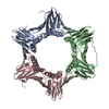
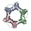
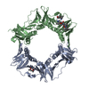
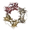
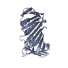
Title
Crystal Structure of the Human Rad9-Rad1-Hus1 DNA Damage Checkpoint Complex
Components
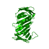
Cell cycle checkpoint control protein RAD9A
Cell cycle checkpoint protein RAD1
Checkpoint protein HUS1
Keywords
CELL CYCLE / PCNA / DNA BINDING CLAMP / DNA damage / DNA repair / Exonuclease / Hydrolase / Nuclease / Nucleus / Phosphoprotein
Function / homology
Function and homology information
meiotic DNA integrity checkpoint signaling / checkpoint clamp complex / meiotic recombination checkpoint signaling / double-stranded DNA 3'-5' DNA exonuclease activity / exodeoxyribonuclease III / DNA replication checkpoint signaling / embryo development ending in birth or egg hatching / positive regulation of intrinsic apoptotic signaling pathway in response to DNA damage / mitotic DNA replication checkpoint signaling / mitotic intra-S DNA damage checkpoint signaling ...meiotic DNA integrity checkpoint signaling / checkpoint clamp complex / meiotic recombination checkpoint signaling / double-stranded DNA 3'-5' DNA exonuclease activity / exodeoxyribonuclease III / DNA replication checkpoint signaling / embryo development ending in birth or egg hatching / positive regulation of intrinsic apoptotic signaling pathway in response to DNA damage / mitotic DNA replication checkpoint signaling / mitotic intra-S DNA damage checkpoint signaling / HDR through Single Strand Annealing (SSA) / Impaired BRCA2 binding to RAD51 / Presynaptic phase of homologous DNA pairing and strand exchange / Activation of ATR in response to replication stress / response to UV / 3'-5' exonuclease activity / substantia nigra development / cellular response to ionizing radiation / telomere maintenance / DNA damage checkpoint signaling / double-strand break repair via homologous recombination / nucleotide-excision repair / G2/M DNA damage checkpoint / SH3 domain binding / histone deacetylase binding / intrinsic apoptotic signaling pathway in response to DNA damage / site of double-strand break / Processing of DNA double-strand break ends / chromosome / Regulation of TP53 Activity through Phosphorylation / damaged DNA binding / DNA repair / intracellular membrane-bounded organelle / DNA damage response / protein kinase binding / nucleolus / enzyme binding / nucleoplasm / nucleus / cytosol / cytoplasm Similarity search - Function
In the structure databanks used in Yorodumi, some data are registered as the other names, "COVID-19 virus" and "2019-nCoV". Here are the details of the virus and the list of structure data.
Jan 31, 2019. EMDB accession codes are about to change! (news from PDBe EMDB page)
EMDB accession codes are about to change! (news from PDBe EMDB page)
The allocation of 4 digits for EMDB accession codes will soon come to an end. Whilst these codes will remain in use, new EMDB accession codes will include an additional digit and will expand incrementally as the available range of codes is exhausted. The current 4-digit format prefixed with “EMD-” (i.e. EMD-XXXX) will advance to a 5-digit format (i.e. EMD-XXXXX), and so on. It is currently estimated that the 4-digit codes will be depleted around Spring 2019, at which point the 5-digit format will come into force.
The EM Navigator/Yorodumi systems omit the EMD- prefix.
Related info.:Q: What is EMD? / ID/Accession-code notation in Yorodumi/EM Navigator
Yorodumi is a browser for structure data from EMDB, PDB, SASBDB, etc.
This page is also the successor to EM Navigator detail page, and also detail information page/front-end page for Omokage search.
The word "yorodu" (or yorozu) is an old Japanese word meaning "ten thousand". "mi" (miru) is to see.
Related info.:EMDB / PDB / SASBDB / Comparison of 3 databanks / Yorodumi Search / Aug 31, 2016. New EM Navigator & Yorodumi / Yorodumi Papers / Jmol/JSmol / Function and homology information / Changes in new EM Navigator and Yorodumi
 Movie
Movie Controller

 Yorodumi
 Open data
 Basic information
 Components
 Keywords
 Function and homology information
 Homo sapiens (human)
 X-RAY DIFFRACTION /
  Authors
 Citation
 Structure visualization
 Downloads & links
 Other downloads


 PDBj
PDBj


 Assembly

 Spodoptera frugiperda (fall armyworm) / Strain (production host): SF9 / References: UniProt: Q99638, exodeoxyribonuclease III
 Mass: 18.015 Da / Num. of mol.: 82 / Source method: isolated from a natural source / Formula: H2O
 Sample preparation
 Processing