 Movie
Movie Controller

[English] 日本語
 Yorodumi
- PDB-3g3q: Crystal structure of a eukaryotic polyphosphate polymerase in com... -
+ Open data

- Basic information
| Entry | Database: PDB / ID: 3g3q | ||||||
|---|---|---|---|---|---|---|---|
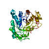
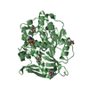
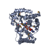
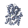
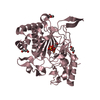
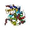
| Title | Crystal structure of a eukaryotic polyphosphate polymerase in complex with a phosphate polymer | ||||||
 Components Components | Vacuolar transporter chaperone 4 | ||||||
 Keywords Keywords | BIOSYNTHETIC PROTEIN / polyphosphate polymerase / polyphosphate kinase / VTC complex / vacuolar transporter chaperone / tunnel enzyme / Membrane / Phosphoprotein / Transmembrane / Vacuole | ||||||
| Function / homology |  Function and homology information Function and homology informationvacuolar transporter chaperone complex / engulfment of target by autophagosome / ATP-polyphosphate phosphotransferase / polyphosphate biosynthetic process / polyphosphate kinase activity / microautophagy / polyphosphate metabolic process / vacuolar transport / fungal-type vacuole membrane / inositol hexakisphosphate binding ...vacuolar transporter chaperone complex / engulfment of target by autophagosome / ATP-polyphosphate phosphotransferase / polyphosphate biosynthetic process / polyphosphate kinase activity / microautophagy / polyphosphate metabolic process / vacuolar transport / fungal-type vacuole membrane / inositol hexakisphosphate binding / vacuolar membrane / autophagosome membrane / cell periphery / cytoplasmic vesicle / cell cortex / calmodulin binding / endoplasmic reticulum membrane / endoplasmic reticulum Similarity search - Function | ||||||
| Biological species |  | ||||||
| Method |  X-RAY DIFFRACTION / SYNCHROTRON / MOLECULAR REPLACEMENT / Resolution: 2.64 Å X-RAY DIFFRACTION / SYNCHROTRON / MOLECULAR REPLACEMENT / Resolution: 2.64 Å | ||||||
 Authors Authors | Hothorn, M. / Scheffzek, K. | ||||||
 Citation Citation | Journal: Science / Year: 2009 Title: Catalytic core of a membrane-associated eukaryotic polyphosphate polymerase. Authors: Hothorn, M. / Neumann, H. / Lenherr, E.D. / Wehner, M. / Rybin, V. / Hassa, P.O. / Uttenweiler, A. / Reinhardt, M. / Schmidt, A. / Seiler, J. / Ladurner, A.G. / Herrmann, C. / Scheffzek, K. / Mayer, A. | ||||||
| History |
|
- Structure visualization
| Structure viewer | Molecule: MolmilJmol/JSmol |
|---|
- Downloads & links
-Download
| PDBx/mmCIF format | 3g3q.cif.gz | 136.9 KB | Display | PDBx/mmCIF format |
|---|---|---|---|---|
| PDB format | pdb3g3q.ent.gz | 106.9 KB | Display | PDB format |
| PDBx/mmJSON format | 3g3q.json.gz | Tree view | PDBx/mmJSON format | |
| Others |  Other downloads Other downloads |
-Validation report
| Arichive directory | https://data.pdbj.org/pub/pdb/validation_reports/g3/3g3qftp://data.pdbj.org/pub/pdb/validation_reports/g3/3g3q | HTTPS FTP |
|---|
-Related structure data
| Related structure data |  3g3oSC  3g3rC  3g3tC  3g3uC S: Starting model for refinement C: citing same article ( |
|---|---|
| Similar structure data |


-Links
 PDBj
PDBj- Assembly
| Deposited unit | 
| ||||||||||||||||||
|---|---|---|---|---|---|---|---|---|---|---|---|---|---|---|---|---|---|---|---|
| 1 | 
| ||||||||||||||||||
| 2 | 
| ||||||||||||||||||
| 3 |
| ||||||||||||||||||
| Unit cell |
| ||||||||||||||||||
| Noncrystallographic symmetry (NCS) | NCS domain:
NCS domain segments:
|
-Components
| #1: Protein | Mass: 34773.754 Da / Num. of mol.: 2 / Fragment: UNP residues 189-480 Source method: isolated from a genetically manipulated source Source: (gene. exp.) Gene: J1345, PHM3, VTC4, YJL012C / Plasmid: pETM11 / Production host:  #2: Chemical | ChemComp-PO4 /   Mass: 94.971 Da / Num. of mol.: 29 / Source method: obtained synthetically / Formula: PO4 Mass: 94.971 Da / Num. of mol.: 29 / Source method: obtained synthetically / Formula: PO4#3: Chemical |   Mass: 96.063 Da / Num. of mol.: 3 / Source method: obtained synthetically / Formula: SO4 Mass: 96.063 Da / Num. of mol.: 3 / Source method: obtained synthetically / Formula: SO4#4: Water | ChemComp-HOH / |  Mass: 18.015 Da / Num. of mol.: 57 / Source method: isolated from a natural source / Formula: H2O Mass: 18.015 Da / Num. of mol.: 57 / Source method: isolated from a natural source / Formula: H2ONonpolymer details | THERE ARE 29 ORDERED PO4 UNITS IN THE STRUCTURE. THIS POLYPHOSPHATE CHAIN IS SEVERAL HUNDRED UNITS ...THERE ARE 29 ORDERED PO4 UNITS IN THE STRUCTURE. THIS POLYPHOSPH | |
|---|
-Experimental details
-Experiment
| Experiment | Method: X-RAY DIFFRACTION / Number of used crystals: 1 |
|---|
- Sample preparation
| Crystal | Density Matthews: 4.06 Å3/Da / Density % sol: 69.72 % |
|---|---|
| Crystal grow | Temperature: 294 K / Method: vapor diffusion, sitting drop / pH: 7.5 Details: 22% PEG 3350, 0.15 M (NH4)2SO4, 0.1 M Bis-Tris, pH 7.5, VAPOR DIFFUSION, SITTING DROP, temperature 294K |
-Data collection
| Diffraction | Mean temperature: 100 K |
|---|---|
| Diffraction source | Source: SYNCHROTRON / Site: ESRF  / Beamline: BM14 / Wavelength: 0.976 Å / Beamline: BM14 / Wavelength: 0.976 Å |
| Detector | Type: MARMOSAIC 225 mm CCD / Detector: CCD / Date: Apr 12, 2007 |
| Radiation | Monochromator: Si(111) monochromator / Protocol: SINGLE WAVELENGTH / Monochromatic (M) / Laue (L): M / Scattering type: x-ray |
| Radiation wavelength | Wavelength: 0.976 Å / Relative weight: 1 |
| Reflection | Resolution: 2.64→20 Å / Num. all: 32041 / Num. obs: 32041 / % possible obs: 99.7 % / Observed criterion σ(F): -3 / Observed criterion σ(I): -3 / Redundancy: 3.9 % / Biso Wilson estimate: 78.4 Å2 / Rsym value: 0.044 / Net I/σ(I): 18.5 |
| Reflection shell | Resolution: 2.64→2.8 Å / Redundancy: 3.8 % / Mean I/σ(I) obs: 2 / Num. unique all: 5063 / Rsym value: 0.708 / % possible all: 99.6 |
- Processing
| Software |
| |||||||||||||||||||||||||||||||||||||||||||||||||||||||||||||||||||||||||||||||||||||||||||
|---|---|---|---|---|---|---|---|---|---|---|---|---|---|---|---|---|---|---|---|---|---|---|---|---|---|---|---|---|---|---|---|---|---|---|---|---|---|---|---|---|---|---|---|---|---|---|---|---|---|---|---|---|---|---|---|---|---|---|---|---|---|---|---|---|---|---|---|---|---|---|---|---|---|---|---|---|---|---|---|---|---|---|---|---|---|---|---|---|---|---|---|---|
| Refinement | Method to determine structure: MOLECULAR REPLACEMENT Starting model: PDB entry 3g3o Resolution: 2.64→19.824 Å / SU ML: 0.43 / σ(F): 1.97 / Stereochemistry target values: ML
| |||||||||||||||||||||||||||||||||||||||||||||||||||||||||||||||||||||||||||||||||||||||||||
| Solvent computation | Shrinkage radii: 0.9 Å / VDW probe radii: 1.11 Å / Solvent model: FLAT BULK SOLVENT MODEL / Bsol: 59.033 Å2 / ksol: 0.315 e/Å3 | |||||||||||||||||||||||||||||||||||||||||||||||||||||||||||||||||||||||||||||||||||||||||||
| Refinement step | Cycle: LAST / Resolution: 2.64→19.824 Å
| |||||||||||||||||||||||||||||||||||||||||||||||||||||||||||||||||||||||||||||||||||||||||||
| Refine LS restraints |
| |||||||||||||||||||||||||||||||||||||||||||||||||||||||||||||||||||||||||||||||||||||||||||
| Refine LS restraints NCS |
| |||||||||||||||||||||||||||||||||||||||||||||||||||||||||||||||||||||||||||||||||||||||||||
| LS refinement shell |
|
