 Movie
Movie Controller

[English] 日本語
 Yorodumi
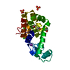
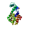
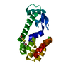
- PDB-3g21: Crystal structure of the C-terminal domain of the Rous Sarcoma Vi... -
+ Open data

- Basic information
| Entry | Database: PDB / ID: 3g21 | ||||||
|---|---|---|---|---|---|---|---|
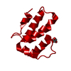
| Title | Crystal structure of the C-terminal domain of the Rous Sarcoma Virus capsid protein: Low pH | ||||||
 Components Components | Gag polyprotein | ||||||
 Keywords Keywords | VIRAL PROTEIN / ALPHA-HELICAL BUNDLE / CAPSID PROTEIN / VIRION / RETROVIRUS | ||||||
| Function / homology |  Function and homology information Function and homology informationhost cell nucleoplasm / viral procapsid maturation / host cell nucleolus / Hydrolases; Acting on peptide bonds (peptidases); Aspartic endopeptidases / viral capsid / aspartic-type endopeptidase activity / structural constituent of virion / nucleic acid binding / viral translational frameshifting / host cell plasma membrane ...host cell nucleoplasm / viral procapsid maturation / host cell nucleolus / Hydrolases; Acting on peptide bonds (peptidases); Aspartic endopeptidases / viral capsid / aspartic-type endopeptidase activity / structural constituent of virion / nucleic acid binding / viral translational frameshifting / host cell plasma membrane / proteolysis / zinc ion binding / membrane Similarity search - Function | ||||||
| Biological species |  Rous sarcoma virus Rous sarcoma virus | ||||||
| Method |  X-RAY DIFFRACTION / SYNCHROTRON / MOLECULAR REPLACEMENT / Resolution: 0.9 Å X-RAY DIFFRACTION / SYNCHROTRON / MOLECULAR REPLACEMENT / Resolution: 0.9 Å | ||||||
 Authors Authors | Kingston, R.L. | ||||||
 Citation Citation | Journal: Structure / Year: 2009 Title: Proton-linked dimerization of a retroviral capsid protein initiates capsid assembly Authors: Bailey, G.D. / Hyun, J.K. / Mitra, A.K. / Kingston, R.L. | ||||||
| History |
|
- Structure visualization
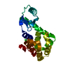
| Structure viewer | Molecule: MolmilJmol/JSmol |
|---|
- Downloads & links
-Download
| PDBx/mmCIF format | 3g21.cif.gz | 51.2 KB | Display | PDBx/mmCIF format |
|---|---|---|---|---|
| PDB format | pdb3g21.ent.gz | 36.7 KB | Display | PDB format |
| PDBx/mmJSON format | 3g21.json.gz | Tree view | PDBx/mmJSON format | |
| Others |  Other downloads Other downloads |
-Validation report
| Summary document | 3g21_validation.pdf.gz | 414.8 KB | Display | wwPDB validaton report |
|---|---|---|---|---|
| Full document | 3g21_full_validation.pdf.gz | 416 KB | Display | |
| Data in XML | 3g21_validation.xml.gz | 6.7 KB | Display | |
| Data in CIF | 3g21_validation.cif.gz | 8.6 KB | Display | |
| Arichive directory | https://data.pdbj.org/pub/pdb/validation_reports/g2/3g21ftp://data.pdbj.org/pub/pdb/validation_reports/g2/3g21 | HTTPS FTP |
-Related structure data
| Related structure data |  3g0vC  3g1gSC  3g1iC  3g26C  3g28C  3g29C C: citing same article ( S: Starting model for refinement |
|---|---|
| Similar structure data |


-Links
 PDBj
PDBj- Assembly
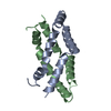
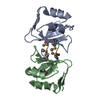
| Deposited unit | 
| ||||||||
|---|---|---|---|---|---|---|---|---|---|
| 1 | 
| ||||||||
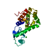
| Unit cell |
|
-Components
| #1: Protein | Mass: 8474.636 Da / Num. of mol.: 1 / Fragment: C-terminal domain, UNP residues 389-465 Source method: isolated from a genetically manipulated source Source: (gene. exp.) Rous sarcoma virus / Strain: Prague C Strain / Gene: gag / Plasmid: pTYB11 / Production host:  References: UniProt: P03322, Hydrolases; Acting on peptide bonds (peptidases); Aspartic endopeptidases |
|---|---|
| #2: Chemical | ChemComp-NO3 /   Mass: 62.005 Da / Num. of mol.: 1 / Source method: obtained synthetically / Formula: NO3 Mass: 62.005 Da / Num. of mol.: 1 / Source method: obtained synthetically / Formula: NO3 |
| #3: Water | ChemComp-HOH /  Mass: 18.015 Da / Num. of mol.: 83 / Source method: isolated from a natural source / Formula: H2O Mass: 18.015 Da / Num. of mol.: 83 / Source method: isolated from a natural source / Formula: H2O |
-Experimental details
-Experiment
| Experiment | Method: X-RAY DIFFRACTION / Number of used crystals: 1 |
|---|
- Sample preparation
| Crystal | Density Matthews: 2.01 Å3/Da / Density % sol: 38.75 % |
|---|---|
| Crystal grow | Temperature: 291 K / Method: vapor diffusion, sitting drop / pH: 4.3 Details: 0.20M Succinic acid/KOH, pH4.3, 13-18% PEG8000, 0.75M Magnesium Nitrate, VAPOR DIFFUSION, SITTING DROP, temperature 291K |
-Data collection
| Diffraction | Mean temperature: 100 K |
|---|---|
| Diffraction source | Source: SYNCHROTRON / Site: SSRL  / Beamline: BL9-2 / Wavelength: 0.85503 Å / Beamline: BL9-2 / Wavelength: 0.85503 Å |
| Detector | Type: MARMOSAIC 325 mm CCD / Detector: CCD / Date: Apr 28, 2008 |
| Radiation | Monochromator: Double crystal monochromator Si(111) / Protocol: SINGLE WAVELENGTH / Monochromatic (M) / Laue (L): M / Scattering type: x-ray |
| Radiation wavelength | Wavelength: 0.85503 Å / Relative weight: 1 |
| Reflection | Resolution: 0.9→27 Å / Num. all: 46299 / Num. obs: 46299 / % possible obs: 89 % / Observed criterion σ(F): 0 / Observed criterion σ(I): 0 / Redundancy: 11.1 % / Rmerge(I) obs: 0.058 / Net I/σ(I): 36.2 |
| Reflection shell | Resolution: 0.9→0.93 Å / Redundancy: 1.8 % / Rmerge(I) obs: 0.167 / Mean I/σ(I) obs: 7.2 / Num. unique all: 1546 / % possible all: 30 |
- Processing
| Software |
| |||||||||||||||||||||||||||||||||
|---|---|---|---|---|---|---|---|---|---|---|---|---|---|---|---|---|---|---|---|---|---|---|---|---|---|---|---|---|---|---|---|---|---|---|
| Refinement | Method to determine structure: MOLECULAR REPLACEMENT Starting model: PDB ENTRY 3G1G Resolution: 0.9→27 Å / Num. parameters: 7348 / Num. restraintsaints: 10719 Isotropic thermal model: Restrained Anisotropic Thermal Parameters Cross valid method: FREE R / σ(F): 0 / σ(I): 0 / Stereochemistry target values: Engh & Huber / Details: ANISOTROPIC REFINEMENT REDUCED FREE R (NO CUTOFF)
| |||||||||||||||||||||||||||||||||
| Solvent computation | Solvent model: Moews and Kretsinger (Babinet scaling) | |||||||||||||||||||||||||||||||||
| Refine analyze | Num. disordered residues: 42 / Occupancy sum hydrogen: 583.31 / Occupancy sum non hydrogen: 669.09 | |||||||||||||||||||||||||||||||||
| Refinement step | Cycle: LAST / Resolution: 0.9→27 Å
| |||||||||||||||||||||||||||||||||
| Refine LS restraints |
|
