 Movie
Movie Controller

[English] 日本語
 Yorodumi
- PDB-3g0v: Crystal structure of the C-terminal domain from the Rous Sarcoma ... -
+ Open data

- Basic information
| Entry | Database: PDB / ID: 3g0v | ||||||
|---|---|---|---|---|---|---|---|
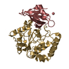
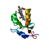
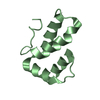
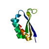
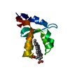
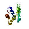
| Title | Crystal structure of the C-terminal domain from the Rous Sarcoma Virus capsid protein: mutant D179A | ||||||
 Components Components | Gag polyprotein | ||||||
 Keywords Keywords | VIRAL PROTEIN / ALPHA-HELICAL BUNDLE / CAPSID PROTEIN / VIRION / RETROVIRUS | ||||||
| Function / homology |  Function and homology information Function and homology informationhost cell nucleoplasm / viral procapsid maturation / host cell nucleolus / Hydrolases; Acting on peptide bonds (peptidases); Aspartic endopeptidases / viral capsid / structural constituent of virion / aspartic-type endopeptidase activity / nucleic acid binding / viral translational frameshifting / host cell plasma membrane ...host cell nucleoplasm / viral procapsid maturation / host cell nucleolus / Hydrolases; Acting on peptide bonds (peptidases); Aspartic endopeptidases / viral capsid / structural constituent of virion / aspartic-type endopeptidase activity / nucleic acid binding / viral translational frameshifting / host cell plasma membrane / proteolysis / zinc ion binding Similarity search - Function | ||||||
| Biological species |  Rous sarcoma virus Rous sarcoma virus | ||||||
| Method |  X-RAY DIFFRACTION / MOLECULAR REPLACEMENT / Resolution: 2 Å X-RAY DIFFRACTION / MOLECULAR REPLACEMENT / Resolution: 2 Å | ||||||
 Authors Authors | Kingston, R.L. | ||||||
 Citation Citation | Journal: Structure / Year: 2009 Title: Proton-linked dimerization of a retroviral capsid protein initiates capsid assembly Authors: Bailey, G.D. / Hyun, J.K. / Mitra, A.K. / Kingston, R.L. | ||||||
| History |
|
- Structure visualization
| Structure viewer | Molecule: MolmilJmol/JSmol |
|---|
- Downloads & links
-Download
| PDBx/mmCIF format | 3g0v.cif.gz | 28.1 KB | Display | PDBx/mmCIF format |
|---|---|---|---|---|
| PDB format | pdb3g0v.ent.gz | 17.9 KB | Display | PDB format |
| PDBx/mmJSON format | 3g0v.json.gz | Tree view | PDBx/mmJSON format | |
| Others |  Other downloads Other downloads |
-Validation report
| Arichive directory | https://data.pdbj.org/pub/pdb/validation_reports/g0/3g0vftp://data.pdbj.org/pub/pdb/validation_reports/g0/3g0v | HTTPS FTP |
|---|
-Related structure data
| Related structure data |  3g1gC  3g1iC 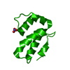 3g21C  3g26C  3g28SC  3g29C C: citing same article ( S: Starting model for refinement |
|---|---|
| Similar structure data |


-Links
 PDBj
PDBj- Assembly
| Deposited unit | 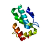
| ||||||||
|---|---|---|---|---|---|---|---|---|---|
| 1 |
| ||||||||
| Unit cell |
| ||||||||
| Components on special symmetry positions |
|
-Components
| #1: Protein | Mass: 8430.626 Da / Num. of mol.: 1 / Fragment: C-terminal domain, UNP residues 389-465 / Mutation: D179A Source method: isolated from a genetically manipulated source Source: (gene. exp.) Rous sarcoma virus / Strain: Prague C Strain / Gene: gag / Plasmid: pTYB11 / Production host:  References: UniProt: P03322, Hydrolases; Acting on peptide bonds (peptidases); Aspartic endopeptidases |
|---|---|
| #2: Chemical | ChemComp-NO3 /   Mass: 62.005 Da / Num. of mol.: 1 / Source method: obtained synthetically / Formula: NO3 Mass: 62.005 Da / Num. of mol.: 1 / Source method: obtained synthetically / Formula: NO3 |
| #3: Water | ChemComp-HOH /  Mass: 18.015 Da / Num. of mol.: 49 / Source method: isolated from a natural source / Formula: H2O Mass: 18.015 Da / Num. of mol.: 49 / Source method: isolated from a natural source / Formula: H2O |
-Experimental details
-Experiment
| Experiment | Method: X-RAY DIFFRACTION / Number of used crystals: 1 |
|---|
- Sample preparation
| Crystal | Density Matthews: 2.69 Å3/Da / Density % sol: 54.29 % |
|---|---|
| Crystal grow | Temperature: 291 K / Method: vapor diffusion, sitting drop / pH: 4.3 Details: 0.2M Succinic acid/KOH, pH4.3, 24% PEG8000, 1M Sodium Nitrate, VAPOR DIFFUSION, SITTING DROP, temperature 291K |
-Data collection
| Diffraction | Mean temperature: 110 K |
|---|---|
| Diffraction source | Source: ROTATING ANODE / Type: RIGAKU MICROMAX-007 HF / Wavelength: 1.54178 Å |
| Detector | Type: MAR scanner 345 mm plate / Detector: IMAGE PLATE / Date: Oct 21, 2008 / Details: Rigaku Varimax HF confocal optics |
| Radiation | Monochromator: Rigaku Varimax HF confocal optics / Protocol: SINGLE WAVELENGTH / Monochromatic (M) / Laue (L): M / Scattering type: x-ray |
| Radiation wavelength | Wavelength: 1.54178 Å / Relative weight: 1 |
| Reflection | Resolution: 2→38 Å / Num. all: 6413 / Num. obs: 6413 / % possible obs: 100 % / Observed criterion σ(F): 0 / Observed criterion σ(I): 0 / Redundancy: 10.7 % / Rmerge(I) obs: 0.078 / Net I/σ(I): 9.2 |
| Reflection shell | Resolution: 2→2.07 Å / Redundancy: 10.5 % / Rmerge(I) obs: 0.333 / Mean I/σ(I) obs: 2.8 / Num. unique all: 630 / % possible all: 100 |
- Processing
| Software |
| ||||||||||||||||||||||||||||||||||||||||||||||||||||||||||||||||||||||||||||||||||||||||||||||||||||||||||||||||||||||||||||||||||||||||||||||||||||||||||||||||||||||||||
|---|---|---|---|---|---|---|---|---|---|---|---|---|---|---|---|---|---|---|---|---|---|---|---|---|---|---|---|---|---|---|---|---|---|---|---|---|---|---|---|---|---|---|---|---|---|---|---|---|---|---|---|---|---|---|---|---|---|---|---|---|---|---|---|---|---|---|---|---|---|---|---|---|---|---|---|---|---|---|---|---|---|---|---|---|---|---|---|---|---|---|---|---|---|---|---|---|---|---|---|---|---|---|---|---|---|---|---|---|---|---|---|---|---|---|---|---|---|---|---|---|---|---|---|---|---|---|---|---|---|---|---|---|---|---|---|---|---|---|---|---|---|---|---|---|---|---|---|---|---|---|---|---|---|---|---|---|---|---|---|---|---|---|---|---|---|---|---|---|---|---|---|
| Refinement | Method to determine structure: MOLECULAR REPLACEMENT Starting model: PDB ENTRY 3G28 Resolution: 2→38 Å / Cor.coef. Fo:Fc: 0.942 / Cor.coef. Fo:Fc free: 0.923 / SU B: 3.499 / SU ML: 0.097 / Isotropic thermal model: Isotropic / Cross valid method: THROUGHOUT / σ(F): 0 / σ(I): 0 / ESU R: 0.174 / ESU R Free: 0.153 / Stereochemistry target values: MAXIMUM LIKELIHOOD
| ||||||||||||||||||||||||||||||||||||||||||||||||||||||||||||||||||||||||||||||||||||||||||||||||||||||||||||||||||||||||||||||||||||||||||||||||||||||||||||||||||||||||||
| Solvent computation | Ion probe radii: 0.8 Å / Shrinkage radii: 0.8 Å / VDW probe radii: 1.4 Å / Solvent model: MASK | ||||||||||||||||||||||||||||||||||||||||||||||||||||||||||||||||||||||||||||||||||||||||||||||||||||||||||||||||||||||||||||||||||||||||||||||||||||||||||||||||||||||||||
| Displacement parameters | Biso mean: 21.479 Å2
| ||||||||||||||||||||||||||||||||||||||||||||||||||||||||||||||||||||||||||||||||||||||||||||||||||||||||||||||||||||||||||||||||||||||||||||||||||||||||||||||||||||||||||
| Refinement step | Cycle: LAST / Resolution: 2→38 Å
| ||||||||||||||||||||||||||||||||||||||||||||||||||||||||||||||||||||||||||||||||||||||||||||||||||||||||||||||||||||||||||||||||||||||||||||||||||||||||||||||||||||||||||
| Refine LS restraints |
| ||||||||||||||||||||||||||||||||||||||||||||||||||||||||||||||||||||||||||||||||||||||||||||||||||||||||||||||||||||||||||||||||||||||||||||||||||||||||||||||||||||||||||
| LS refinement shell | Resolution: 2.004→2.056 Å / Total num. of bins used: 20
|
