 Movie
Movie Controller

[English] 日本語
 Yorodumi
- PDB-3g02: Structure of enantioselective mutant of epoxide hydrolase from As... -
+ Open data

- Basic information
| Entry | Database: PDB / ID: 3g02 | ||||||
|---|---|---|---|---|---|---|---|
| Title | Structure of enantioselective mutant of epoxide hydrolase from Aspergillus niger generated by directed evolution | ||||||
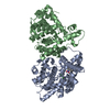
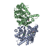
 Components Components | Epoxide hydrolase | ||||||
 Keywords Keywords | HYDROLASE / EPOXIDE HYDROLASE / ALPHA/BETA HYDROLASE FOLD / enantioselective / mutant / directed evolution | ||||||
| Function / homology |  Function and homology information Function and homology informationepoxide hydrolase / epoxide metabolic process / epoxide hydrolase activity Similarity search - Function | ||||||
| Biological species |  | ||||||
| Method |  X-RAY DIFFRACTION / SYNCHROTRON / MOLECULAR REPLACEMENT / molecular replacement / Resolution: 1.5 Å X-RAY DIFFRACTION / SYNCHROTRON / MOLECULAR REPLACEMENT / molecular replacement / Resolution: 1.5 Å | ||||||
 Authors Authors | Naworyta, A. / Mowbray, S.L. | ||||||
 Citation Citation | Journal: J.Am.Chem.Soc. / Year: 2009 Title: Directed evolution of an enantioselective epoxide hydrolase: uncovering the source of enantioselectivity at each evolutionary stage Authors: Reetz, M.T. / Bocola, M. / Wang, L.-W. / Sanchis, J. / Cronin, A. / Arand, M. / Zou, J. / Archelas, A. / Bottalla, A.-L. / Naworyta, A. / Mowbray, S.L. #1: Journal: Structure / Year: 2000Title: Structure of Aspergillus niger epoxide hydrolase at 1.8 A resolution: implications for the structure and function of the mammalian microsomal class of epoxide hydrolases Authors: Zou, J. / Hallberg, B.M. / Bergfors, T. / Oesch, F. / Arand, M. / Mowbray, S.L. / Jones, T.A. | ||||||
| History |
|
- Structure visualization
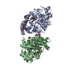
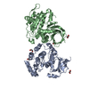
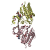
| Structure viewer | Molecule: MolmilJmol/JSmol |
|---|
- Downloads & links
-Download
| PDBx/mmCIF format | 3g02.cif.gz | 181.9 KB | Display | PDBx/mmCIF format |
|---|---|---|---|---|
| PDB format | pdb3g02.ent.gz | 143 KB | Display | PDB format |
| PDBx/mmJSON format | 3g02.json.gz | Tree view | PDBx/mmJSON format | |
| Others |  Other downloads Other downloads |
-Validation report
| Arichive directory | https://data.pdbj.org/pub/pdb/validation_reports/g0/3g02ftp://data.pdbj.org/pub/pdb/validation_reports/g0/3g02 | HTTPS FTP |
|---|
-Related structure data
| Related structure data | 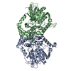 3g0iC 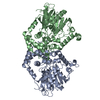 1qo7S S: Starting model for refinement C: citing same article ( |
|---|---|
| Similar structure data |


-Links
 PDBj
PDBj

- Assembly
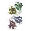
| Deposited unit | 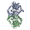
| ||||||||
|---|---|---|---|---|---|---|---|---|---|
| 1 |
| ||||||||
| Unit cell |
|
-Components
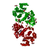
| #1: Protein | Mass: 46047.125 Da / Num. of mol.: 2 / Fragment: residues 5-398 Mutation: L215F, A217N, R219S, L249Y, T317W, T318V, M329P, L330Y, C350V Source method: isolated from a genetically manipulated source Source: (gene. exp.)  #2: Chemical |   Mass: 46.025 Da / Num. of mol.: 3 / Source method: obtained synthetically / Formula: CH2O2 Mass: 46.025 Da / Num. of mol.: 3 / Source method: obtained synthetically / Formula: CH2O2#3: Water | ChemComp-HOH / |  Mass: 18.015 Da / Num. of mol.: 706 / Source method: isolated from a natural source / Formula: H2O Mass: 18.015 Da / Num. of mol.: 706 / Source method: isolated from a natural source / Formula: H2O |
|---|
-Experimental details
-Experiment
| Experiment | Method: X-RAY DIFFRACTION / Number of used crystals: 1 |
|---|
- Sample preparation
| Crystal | Density Matthews: 2.19 Å3/Da / Density % sol: 43.94 % |
|---|---|
| Crystal grow | Temperature: 293 K / Method: vapor diffusion, sitting drop / pH: 6 Details: 20% PEG 6000, 0.1M MES, pH 6.0, 0.1M unbuffered sodium acetate, VAPOR DIFFUSION, SITTING DROP, temperature 293K |
-Data collection
| Diffraction | Mean temperature: 100 K |
|---|---|
| Diffraction source | Source: SYNCHROTRON / Site: ESRF  / Beamline: ID14-2 / Wavelength: 0.933 Å / Beamline: ID14-2 / Wavelength: 0.933 Å |
| Detector | Type: ADSC QUANTUM 4 / Detector: CCD / Date: Aug 31, 2007 |
| Radiation | Monochromator: graphite / Protocol: SINGLE WAVELENGTH / Monochromatic (M) / Laue (L): M / Scattering type: x-ray |
| Radiation wavelength | Wavelength: 0.933 Å / Relative weight: 1 |
| Reflection | Resolution: 1.5→30.54 Å / Num. all: 209428 / Num. obs: 120696 / % possible obs: 95.5 % / Observed criterion σ(I): 0 / Redundancy: 1.7 % / Biso Wilson estimate: 0 Å2 / Rmerge(I) obs: 0.027 / Net I/σ(I): 15.8 |
| Reflection shell | Resolution: 1.5→1.58 Å / Redundancy: 1.7 % / Rmerge(I) obs: 0.128 / Mean I/σ(I) obs: 7.2 / Num. unique all: 18142 / % possible all: 98.2 |
-Phasing
| Phasing | Method: molecular replacement | |||||||||
|---|---|---|---|---|---|---|---|---|---|---|
| Phasing MR | Model details: Phaser MODE: MR_AUTO
|
- Processing
| Software |
| ||||||||||||||||||||||||||||||||||||||||||||||||||||||||||||||||||||||||||||||||||||||||||
|---|---|---|---|---|---|---|---|---|---|---|---|---|---|---|---|---|---|---|---|---|---|---|---|---|---|---|---|---|---|---|---|---|---|---|---|---|---|---|---|---|---|---|---|---|---|---|---|---|---|---|---|---|---|---|---|---|---|---|---|---|---|---|---|---|---|---|---|---|---|---|---|---|---|---|---|---|---|---|---|---|---|---|---|---|---|---|---|---|---|---|---|
| Refinement | Method to determine structure: MOLECULAR REPLACEMENT Starting model: PDB ENTRY 1QO7 Resolution: 1.5→28.91 Å / Cor.coef. Fo:Fc: 0.956 / Cor.coef. Fo:Fc free: 0.946 / WRfactor Rfree: 0.225 / WRfactor Rwork: 0.207 / Occupancy max: 1 / Occupancy min: 0.25 / FOM work R set: 0.877 / SU B: 1.201 / SU ML: 0.046 / SU R Cruickshank DPI: 0.082 / SU Rfree: 0.079 / Isotropic thermal model: isotropic / Cross valid method: THROUGHOUT / σ(F): 0 / ESU R: 0.081 / ESU R Free: 0.079 / Stereochemistry target values: MAXIMUM LIKELIHOOD / Details: HYDROGENS HAVE BEEN ADDED IN THE RIDING POSITIONS
| ||||||||||||||||||||||||||||||||||||||||||||||||||||||||||||||||||||||||||||||||||||||||||
| Solvent computation | Ion probe radii: 0.8 Å / Shrinkage radii: 0.8 Å / VDW probe radii: 1.4 Å / Solvent model: MASK | ||||||||||||||||||||||||||||||||||||||||||||||||||||||||||||||||||||||||||||||||||||||||||
| Displacement parameters | Biso max: 48.51 Å2 / Biso mean: 15.23 Å2 / Biso min: 3.91 Å2
| ||||||||||||||||||||||||||||||||||||||||||||||||||||||||||||||||||||||||||||||||||||||||||
| Refinement step | Cycle: LAST / Resolution: 1.5→28.91 Å
| ||||||||||||||||||||||||||||||||||||||||||||||||||||||||||||||||||||||||||||||||||||||||||
| Refine LS restraints |
| ||||||||||||||||||||||||||||||||||||||||||||||||||||||||||||||||||||||||||||||||||||||||||
| LS refinement shell | Resolution: 1.5→1.539 Å / Total num. of bins used: 20
|
