[English] 日本語
Yorodumi
- PDB-3fzb: Crystal structure of the tail terminator protein from phage lambd... -

+
Open data


ID or keywords:

Loading...

-
Basic information

Entry
Database: PDB / ID: 3fzb
Title
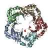
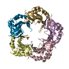
Crystal structure of the tail terminator protein from phage lambda (gpU-WT)
ComponentsMinor tail protein U
KeywordsVIRAL PROTEIN / Mixed Alpha-Beta fold
Function / homology
Function and homology information


symbiont genome ejection through host cell envelope, long flexible tail mechanism / viral tail assembly / virus tail / host cell cytoplasm
Similarity search - Function
Phage minor tail protein U / Minor tail protein U-like / GpU-like superfamily / Phage minor tail protein U / Phage tail protein-like superfamily / Alpha-Beta Plaits / 2-Layer Sandwich / Alpha Beta
Similarity search - Domain/homology
Tail tube terminator protein
Similarity search - Component
Biological speciesEnterobacteria phage lambda (virus)
MethodX-RAY DIFFRACTION / MOLECULAR REPLACEMENT / Resolution: 2.8 Å
AuthorsPell, L.G. / Liu, A. / Edmonds, E. / Donaldson, L.W. / Howell, P.L. / Davidson, A.R.
CitationJournal: J.Mol.Biol. / Year: 2009
Title: The X-ray crystal structure of the phage lambda tail terminator protein reveals the biologically relevant hexameric ring structure and demonstrates a conserved mechanism of tail termination ...Title: The X-ray crystal structure of the phage lambda tail terminator protein reveals the biologically relevant hexameric ring structure and demonstrates a conserved mechanism of tail termination among diverse long-tailed phages.
Authors: Pell, L.G. / Liu, A. / Edmonds, L. / Donaldson, L.W. / Howell, P.L. / Davidson, A.R.
History
DepositionJan 24, 2009Deposition site: RCSB / Processing site: RCSB
Revision 1.0May 26, 2009Provider: repository / Type: Initial release
Revision 1.1Jul 13, 2011Group: Version format compliance
Revision 1.2Jan 22, 2020Group: Advisory / Data collection ...Advisory / Data collection / Database references / Derived calculations
Category: pdbx_struct_assembly / pdbx_struct_assembly_gen ...pdbx_struct_assembly / pdbx_struct_assembly_gen / pdbx_struct_assembly_prop / pdbx_unobs_or_zero_occ_atoms / struct_biol / struct_ref_seq_dif
Item: _struct_ref_seq_dif.details
Revision 1.3Feb 21, 2024Group: Advisory / Data collection ...Advisory / Data collection / Database references / Derived calculations
Category: chem_comp_atom / chem_comp_bond ...chem_comp_atom / chem_comp_bond / database_2 / pdbx_unobs_or_zero_occ_atoms / struct_site
Item: _database_2.pdbx_DOI / _database_2.pdbx_database_accession ..._database_2.pdbx_DOI / _database_2.pdbx_database_accession / _struct_site.pdbx_auth_asym_id / _struct_site.pdbx_auth_comp_id / _struct_site.pdbx_auth_seq_id

-
Structure visualization

Structure viewerMolecule:
MolmilJmol/JSmol

Downloads & links

-
Assembly

Deposited unit
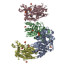
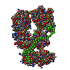
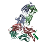
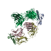
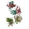
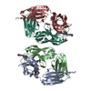
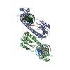
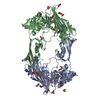
A: Minor tail protein U
B: Minor tail protein U
C: Minor tail protein U
D: Minor tail protein U
E: Minor tail protein U
F: Minor tail protein U
G: Minor tail protein U
H: Minor tail protein U
I: Minor tail protein U
J: Minor tail protein U
hetero molecules


Theoretical massNumber of molelcules
Total (without water)149,70213
Polymers149,41410
Non-polymers2883
Water1,964109
1
A: Minor tail protein U
B: Minor tail protein U
F: Minor tail protein U
H: Minor tail protein U
I: Minor tail protein U
hetero molecules


Theoretical massNumber of molelcules
Total (without water)74,8997
Polymers74,7075
Non-polymers1922
Water905
TypeNameSymmetry operationNumber
identity operation1_555x,y,z1
Buried area6050 Å2
ΔGint-42 kcal/mol
Surface area27390 Å2
MethodPISA
2
C: Minor tail protein U
D: Minor tail protein U
E: Minor tail protein U
G: Minor tail protein U
J: Minor tail protein U
hetero molecules


Theoretical massNumber of molelcules
Total (without water)74,8036
Polymers74,7075
Non-polymers961
Water905
TypeNameSymmetry operationNumber
identity operation1_555x,y,z1
Buried area6060 Å2
ΔGint-37 kcal/mol
Surface area27790 Å2
MethodPISA
Unit cell
Length a, b, c (Å)74.100, 127.990, 82.910
Angle α, β, γ (deg.)90.00, 108.92, 90.00
Int Tables number4
Space group name H-MP1211

-
Components

#1: Protein
Minor tail protein U


Mass: 14941.400 Da / Num. of mol.: 10
Source method: isolated from a genetically manipulated source
Source: (gene. exp.) Enterobacteria phage lambda (virus) / Gene: U / Plasmid: pET15b / Production host: Escherichia coli (E. coli) / Strain (production host): BL21 CodonPlus / References: UniProt: P03732
#2: Chemical ChemComp-SO4 / SULFATE ION


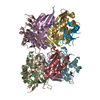
Mass: 96.063 Da / Num. of mol.: 3 / Source method: obtained synthetically / Formula: SO4
#3: Water ChemComp-HOH / water


Mass: 18.015 Da / Num. of mol.: 109 / Source method: isolated from a natural source / Formula: H2O

-
Experimental details

-
Experiment

ExperimentMethod: X-RAY DIFFRACTION / Number of used crystals: 1

-
Sample preparation

CrystalDensity Matthews: 2.49 Å3/Da / Density % sol: 50.59 %
Crystal growTemperature: 298 K / Method: vapor diffusion, hanging drop / pH: 7.5
Details: 0.2M Tri-sodium citrate, 2.2M Ammonium sulfate, pH 7.5, VAPOR DIFFUSION, HANGING DROP, temperature 298K

-
Data collection

DiffractionMean temperature: 100 K
Diffraction sourceSource: ROTATING ANODE / Type: RIGAKU RUH3R / Wavelength: 1.5418 Å
DetectorType: RIGAKU RAXIS / Detector: IMAGE PLATE / Date: Sep 18, 2006
RadiationMonochromator: Osmic optics / Protocol: SINGLE WAVELENGTH / Monochromatic (M) / Laue (L): M / Scattering type: x-ray
Radiation wavelengthWavelength: 1.5418 Å / Relative weight: 1
ReflectionResolution: 2.8→70.1 Å / Num. all: 36045 / Num. obs: 36045 / % possible obs: 99.9 % / Observed criterion σ(F): 0 / Observed criterion σ(I): 0 / Redundancy: 3.93 % / Rmerge(I) obs: 0.099 / Net I/σ(I): 8.6
Reflection shellResolution: 2.8→2.9 Å / Redundancy: 3.93 % / Rmerge(I) obs: 0.312 / Mean I/σ(I) obs: 3.4 / % possible all: 100

-
Processing

Software
NameVersionClassification
CrystalCleardata collection
PHASERphasing
CNS1.1refinement
d*TREKdata reduction
d*TREKdata scaling
RefinementMethod to determine structure: MOLECULAR REPLACEMENT / Resolution: 2.8→70.1 Å / σ(F): 0 / Stereochemistry target values: Engh & Huber
RfactorNum. reflection% reflectionSelection details
Rfree0.309 1788 5 %Random
Rwork0.236 ---
all0.236 36045 --
obs0.236 36045 --
Refinement stepCycle: LAST / Resolution: 2.8→70.1 Å
ProteinNucleic acidLigandSolventTotal
Num. atoms9625 0 15 109 9749
Refine LS restraints
Refine-IDTypeDev ideal
X-RAY DIFFRACTIONc_bond_d0.008
X-RAY DIFFRACTIONc_angle_deg1.5

+
About Yorodumi

-
News

-
Feb 9, 2022. New format data for meta-information of EMDB entries

New format data for meta-information of EMDB entries

  • Version 3 of the EMDB header file is now the official format.
  • The previous official version 1.9 will be removed from the archive.

Related info.:EMDB header

External links:wwPDB to switch to version 3 of the EMDB data model

-
Aug 12, 2020. Covid-19 info

Covid-19 info

URL: https://pdbjlvh1.pdbj.org/emnavi/covid19.php

New page: Covid-19 featured information page in EM Navigator.

Related info.:Covid-19 info / Mar 5, 2020. Novel coronavirus structure data

+
Mar 5, 2020. Novel coronavirus structure data

Novel coronavirus structure data

Related info.:Yorodumi Speices / Aug 12, 2020. Covid-19 info

External links:COVID-19 featured content - PDBj / Molecule of the Month (242):Coronavirus Proteases

+
Jan 31, 2019. EMDB accession codes are about to change! (news from PDBe EMDB page)

EMDB accession codes are about to change! (news from PDBe EMDB page)

  • The allocation of 4 digits for EMDB accession codes will soon come to an end. Whilst these codes will remain in use, new EMDB accession codes will include an additional digit and will expand incrementally as the available range of codes is exhausted. The current 4-digit format prefixed with “EMD-” (i.e. EMD-XXXX) will advance to a 5-digit format (i.e. EMD-XXXXX), and so on. It is currently estimated that the 4-digit codes will be depleted around Spring 2019, at which point the 5-digit format will come into force.
  • The EM Navigator/Yorodumi systems omit the EMD- prefix.

Related info.:Q: What is EMD? / ID/Accession-code notation in Yorodumi/EM Navigator

External links:EMDB Accession Codes are Changing Soon! / Contact to PDBj

+
Jul 12, 2017. Major update of PDB

Major update of PDB

  • wwPDB released updated PDB data conforming to the new PDBx/mmCIF dictionary.
  • This is a major update changing the version number from 4 to 5, and with Remediation, in which all the entries are updated.
  • In this update, many items about electron microscopy experimental information are reorganized (e.g. em_software).
  • Now, EM Navigator and Yorodumi are based on the updated data.

External links:wwPDB Remediation / Enriched Model Files Conforming to OneDep Data Standards Now Available in the PDB FTP Archive

-
Yorodumi

Thousand views of thousand structures

  • Yorodumi is a browser for structure data from EMDB, PDB, SASBDB, etc.
  • This page is also the successor to EM Navigator detail page, and also detail information page/front-end page for Omokage search.
  • The word "yorodu" (or yorozu) is an old Japanese word meaning "ten thousand". "mi" (miru) is to see.

Related info.:EMDB / PDB / SASBDB / Comparison of 3 databanks / Yorodumi Search / Aug 31, 2016. New EM Navigator & Yorodumi / Yorodumi Papers / Jmol/JSmol / Function and homology information / Changes in new EM Navigator and Yorodumi

Read more