 Movie
Movie Controller

[English] 日本語
 Yorodumi
- PDB-3fyr: Crystal structure of the sporulation histidine kinase inhibitor S... -
+ Open data

- Basic information
| Entry | Database: PDB / ID: 3fyr | ||||||
|---|---|---|---|---|---|---|---|
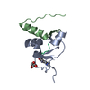
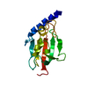
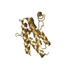
| Title | Crystal structure of the sporulation histidine kinase inhibitor Sda from Bacillus subtilis | ||||||
 Components Components | Sporulation inhibitor sda | ||||||
 Keywords Keywords | Transferase inhibitor / helical hairpin / histidine kinase inhibitor / sporulation regulation / Alternative initiation / Protein kinase inhibitor / Sporulation | ||||||
| Function / homology | Sporulation inhibitor A / Sporulation inhibitor A superfamily / Sporulation inhibitor A / sporulation resulting in formation of a cellular spore / protein kinase inhibitor activity / Sporulation inhibitor sda Function and homology information Function and homology information | ||||||
| Biological species |  | ||||||
| Method |  X-RAY DIFFRACTION / SYNCHROTRON / MAD / Resolution: 1.97 Å X-RAY DIFFRACTION / SYNCHROTRON / MAD / Resolution: 1.97 Å | ||||||
 Authors Authors | Jacques, D.A. / Streamer, M. / King, G.F. / Guss, J.M. / Trewhella, J. / Langley, D.B. | ||||||
 Citation Citation | Journal: Acta Crystallogr.,Sect.D / Year: 2009 Title: Structure of the sporulation histidine kinase inhibitor Sda from Bacillus subtilis and insights into its solution state Authors: Jacques, D.A. / Streamer, M. / Rowland, S.L. / King, G.F. / Guss, J.M. / Trewhella, J. / Langley, D.B. | ||||||
| History |
|
- Structure visualization
| Structure viewer | Molecule: MolmilJmol/JSmol |
|---|
- Downloads & links
-Download
| PDBx/mmCIF format | 3fyr.cif.gz | 34.9 KB | Display | PDBx/mmCIF format |
|---|---|---|---|---|
| PDB format | pdb3fyr.ent.gz | 24.1 KB | Display | PDB format |
| PDBx/mmJSON format | 3fyr.json.gz | Tree view | PDBx/mmJSON format | |
| Others |  Other downloads Other downloads |
-Validation report
| Arichive directory | https://data.pdbj.org/pub/pdb/validation_reports/fy/3fyrftp://data.pdbj.org/pub/pdb/validation_reports/fy/3fyr | HTTPS FTP |
|---|
-Related structure data
| Similar structure data |
|---|


-Links
 PDBj
PDBj- Assembly
| Deposited unit | 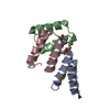
| ||||||||
|---|---|---|---|---|---|---|---|---|---|
| 1 |
| ||||||||
| 2 | 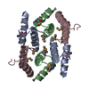
| ||||||||
| Unit cell |
| ||||||||
| Details | THE DEPOSITORS HAVE IN DEPENDANT DATA WHICH SUGGESTS THAT IN SOLUTION THE OLIGOMERIC STATE OF THE ASU (IE AN ODD-LOOKING TRIMER) BEST FITS SAXS DATA OF THE PROTEIN IN SOLUTION. |
-Components
| #1: Protein/peptide | Mass: 5686.198 Da / Num. of mol.: 3 Source method: isolated from a genetically manipulated source Source: (gene. exp.) #2: Water | ChemComp-HOH / |  Mass: 18.015 Da / Num. of mol.: 8 / Source method: isolated from a natural source / Formula: H2O Mass: 18.015 Da / Num. of mol.: 8 / Source method: isolated from a natural source / Formula: H2OHas protein modification | Y | Sequence details | THE FIRST 6 RESIDUES, MNWVPS, ARE MISSING IN NATURAL ACCORDING TO REFERENCE 2, SDA_BACSU IN UNIPROT. ...THE FIRST 6 RESIDUES, MNWVPS, ARE MISSING IN NATURAL ACCORDING TO REFERENCE 2, SDA_BACSU IN UNIPROT. THERE IS AN ALTERNATE START CODON WHICH THE DEPOSITORS | |
|---|
-Experimental details
-Experiment
| Experiment | Method: X-RAY DIFFRACTION / Number of used crystals: 1 |
|---|
- Sample preparation
| Crystal | Density Matthews: 1.718006 Å3/Da / Density % sol: 28.405363 % / Mosaicity: 0.639 ° |
|---|---|
| Crystal grow | Temperature: 293 K / Method: vapor diffusion, hanging drop / pH: 6.3 Details: equal volumes of protein solution (7.5 mg/ml) and well solution (0.1M MES, pH 6.3, 15% (w/v) PEG 5000MME) were combined., VAPOR DIFFUSION, HANGING DROP, temperature 293K |
-Data collection
| Diffraction | Mean temperature: 100 K | |||||||||||||||||||||||||||||||||||||||||||||||||||||||||||||||||||||||||||||
|---|---|---|---|---|---|---|---|---|---|---|---|---|---|---|---|---|---|---|---|---|---|---|---|---|---|---|---|---|---|---|---|---|---|---|---|---|---|---|---|---|---|---|---|---|---|---|---|---|---|---|---|---|---|---|---|---|---|---|---|---|---|---|---|---|---|---|---|---|---|---|---|---|---|---|---|---|---|---|
| Diffraction source | Source: SYNCHROTRON / Site: APS  / Beamline: 23-ID-D / Wavelength: 0.97945, 0.97959, 0.94945 / Beamline: 23-ID-D / Wavelength: 0.97945, 0.97959, 0.94945 | |||||||||||||||||||||||||||||||||||||||||||||||||||||||||||||||||||||||||||||
| Detector | Type: MARMOSAIC 300 mm CCD / Detector: CCD / Date: Feb 20, 2008 | |||||||||||||||||||||||||||||||||||||||||||||||||||||||||||||||||||||||||||||
| Radiation | Protocol: MAD / Monochromatic (M) / Laue (L): M / Scattering type: x-ray | |||||||||||||||||||||||||||||||||||||||||||||||||||||||||||||||||||||||||||||
| Radiation wavelength |
| |||||||||||||||||||||||||||||||||||||||||||||||||||||||||||||||||||||||||||||
| Reflection | Redundancy: 3.6 % / Av σ(I) over netI: 14.87 / Number: 56720 / Rmerge(I) obs: 0.06 / Χ2: 1 / D res high: 1.97 Å / D res low: 50 Å / Num. obs: 15638 / % possible obs: 99.5 | |||||||||||||||||||||||||||||||||||||||||||||||||||||||||||||||||||||||||||||
| Diffraction reflection shell |
| |||||||||||||||||||||||||||||||||||||||||||||||||||||||||||||||||||||||||||||
| Reflection | Resolution: 1.97→50 Å / Num. obs: 8936 / % possible obs: 99.5 % / Redundancy: 3.6 % / Rmerge(I) obs: 0.06 / Χ2: 1.005 / Net I/σ(I): 14.873 | |||||||||||||||||||||||||||||||||||||||||||||||||||||||||||||||||||||||||||||
| Reflection shell | Resolution: 1.97→2.04 Å / Redundancy: 3.1 % / Rmerge(I) obs: 0.564 / Mean I/σ(I) obs: 2 / Num. unique all: 1577 / Χ2: 1.029 / % possible all: 99.9 |
-Phasing
| Phasing | Method: MAD | ||||||||||||||||||||||||||||
|---|---|---|---|---|---|---|---|---|---|---|---|---|---|---|---|---|---|---|---|---|---|---|---|---|---|---|---|---|---|
| Phasing set |
| ||||||||||||||||||||||||||||
| Phasing MAD set |
| ||||||||||||||||||||||||||||
| Phasing MAD set site |
|
- Processing
| Software |
| |||||||||||||||||||||||||||||||||||||||||||||||||||||||||||||||||||||||||||||||||||||
|---|---|---|---|---|---|---|---|---|---|---|---|---|---|---|---|---|---|---|---|---|---|---|---|---|---|---|---|---|---|---|---|---|---|---|---|---|---|---|---|---|---|---|---|---|---|---|---|---|---|---|---|---|---|---|---|---|---|---|---|---|---|---|---|---|---|---|---|---|---|---|---|---|---|---|---|---|---|---|---|---|---|---|---|---|---|---|
| Refinement | Method to determine structure: MAD / Resolution: 1.97→27.69 Å / Cor.coef. Fo:Fc: 0.938 / Cor.coef. Fo:Fc free: 0.879 / WRfactor Rfree: 0.325 / WRfactor Rwork: 0.265 / Occupancy max: 1 / Occupancy min: 1 / FOM work R set: 0.796 / SU B: 4.771 / SU ML: 0.136 / SU R Cruickshank DPI: 0.201 / SU Rfree: 0.198 / Cross valid method: THROUGHOUT / σ(F): 0 / ESU R: 0.201 / ESU R Free: 0.198 / Stereochemistry target values: MAXIMUM LIKELIHOOD Details: HYDROGENS HAVE BEEN ADDED IN THE RIDING POSITIONS; U VALUES : REFINED INDIVIDUALLY
| |||||||||||||||||||||||||||||||||||||||||||||||||||||||||||||||||||||||||||||||||||||
| Solvent computation | Ion probe radii: 0.8 Å / Shrinkage radii: 0.8 Å / VDW probe radii: 1.2 Å / Solvent model: MASK | |||||||||||||||||||||||||||||||||||||||||||||||||||||||||||||||||||||||||||||||||||||
| Displacement parameters | Biso max: 77.98 Å2 / Biso mean: 37.902 Å2 / Biso min: 21.37 Å2
| |||||||||||||||||||||||||||||||||||||||||||||||||||||||||||||||||||||||||||||||||||||
| Refinement step | Cycle: LAST / Resolution: 1.97→27.69 Å
| |||||||||||||||||||||||||||||||||||||||||||||||||||||||||||||||||||||||||||||||||||||
| Refine LS restraints |
| |||||||||||||||||||||||||||||||||||||||||||||||||||||||||||||||||||||||||||||||||||||
| LS refinement shell | Resolution: 1.971→2.022 Å / Total num. of bins used: 20
|
